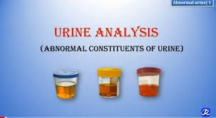
Introduction
- Urine analysis, also called urinalysis, is a commonly used diagnostic laboratory test.
-
It is performed to evaluate the composition of urine.
-
Urinalysis provides important information about overall health, especially the kidneys and urinary system.
-
It is a simple, non-invasive, and cost-effective investigation.
-
A routine urinalysis includes examination of physical, chemical, and microscopic components of urine.
-
Detection of abnormal constituents in urine helps in identifying underlying diseases.
-
Abnormal findings may indicate conditions such as kidney disease, diabetes mellitus, and urinary tract infections.
-
Urine is mainly composed of water, along with small amounts of waste products and electrolytes.
-
Under normal conditions, urine composition remains relatively constant.
-
In pathological states, abnormal substances or altered concentrations of normal components may appear in urine.
Urine collection and preservation
1. Random Specimen
-
The most commonly obtained urine sample, though not the specimen of choice for complete urinalysis.
-
Can be collected at any time of the day.
-
Easy to obtain and readily available.
-
Commonly used for detection of:
-
Glucose
-
Ketone bodies
-
Bile pigments
-
Blood pigments
-
2. Midstream First-Morning Specimen
-
Considered the specimen of choice for routine urinalysis.
-
Collected from the first voided urine in the morning.
-
Urine is more concentrated due to overnight storage in the bladder, increasing diagnostic accuracy.
-
Procedure:
-
The patient discards the initial part of urine.
-
15–20 mL of midstream urine is collected in a clean glass or plastic container.
-
-
Ideal for physical, chemical, and microscopic examination.
3. 24-Hour Urine Collection Specimen
-
Required for quantitative estimation of substances with diurnal variation.
-
Used for measurement of:
-
Creatinine
-
Urea nitrogen
-
Calcium
-
Proteins
-
Glucose
-
Sodium and potassium
-
Hormones (catecholamines, 17-hydroxysteroids, etc.)
-
Procedure:
-
Collection starts at 6:00 AM.
-
The patient empties the bladder and discards the first urine sample.
-
From the second void onward, all urine samples are collected in a container containing preservative.
-
The next day’s 6:00 AM urine sample is collected as the final specimen.
Preservatives Used in Urine Collection
-
Preservatives are mainly used for 24-hour urine collection.
-
Commonly used preservatives include:
-
50 mL of 2 N HCl per 24-hour collection
-
10 mL of concentrated HCl per 24-hour collection
-
Thymol crystals – 5 mL of 100 g/L solution in isopropanol
-
-
Preservatives help prevent:
-
Bacterial growth
-
Decomposition of analytes
-
Sections of Routine Urine Examination
Routine urine examination is carried out under the following three main sections:
-
Physical Examination
-
Includes assessment of:
-
Volume
-
Colour
-
Appearance (clarity/turbidity)
-
Odour
-
Specific gravity
-
pH
-
-
-
Chemical Examination
-
Detects the presence of:
-
Protein
-
Glucose
-
Ketone bodies
-
Bilirubin
-
Urobilinogen
-
Blood
-
-
-
Microscopic Examination
-
Involves identification of:
-
Red blood cells (RBCs)
-
White blood cells (WBCs)
-
Epithelial cells
-
Casts
-
Crystals
-
Microorganisms
-
-
Physical examination
Volume
Normal adult urine output: 1–1.5 litres/day
A. Polyuria (Increased urinary output)
Causes
-
Diabetes mellitus: Osmotic diuresis due to glycosuria
-
Diabetes insipidus: ADH deficiency → inability to concentrate urine
-
Drug-induced diuresis: Digitalis, salicylates, diuretics
-
High fluid intake / psychogenic polydipsia
Clinical Significance
Persistent polyuria suggests renal concentrating defect, endocrine disorders, or osmotic diuresis.
B. Oliguria (Reduced urine output)
Causes
-
Acute nephritis / glomerulonephritis
-
Dehydration: Fever, diarrhoea, vomiting
-
Shock or circulatory collapse
-
Obstruction: Prostate enlargement, ureteric stones
Clinical Significance
Oliguria is a warning sign of acute kidney injury (AKI) or severe volume depletion.
C. Anuria (Total suppression of urine formation)
Causes
-
Shock (severe hypotension → no renal perfusion)
-
Acute nephritis
-
Incompatible blood transfusion reaction
-
Toxins: Mercury poisoning, heavy metals
-
Renal stones blocking both ureters or solitary functioning kidney obstruction
Clinical Significance
Anuria is a medical emergency, suggesting complete renal shutdown or obstruction.

| Parameter | Description / Definition | Clinical Significance |
|---|---|---|
| Normal urine volume | 1–2 liters/day (≈1500 mL) | Indicates normal renal function and hydration |
| Polyuria | >2.5–3 liters/day | Diabetes mellitus, diabetes insipidus, diuretics, excess fluid intake |
| Oliguria | <400 mL/day | Dehydration, shock, acute renal failure |
| Anuria | <100 mL/day | Severe renal failure, urinary tract obstruction |
| Nocturia | Increased urine output at night | Diabetes mellitus, cardiac failure, UTI, prostatic enlargement |
Colour
Normal: Pale yellow due to urochrome pigments
Abnormal Colours & Their Meanings
Smoky brown
-
Indicates hematuria or hemoglobinuria
-
Classic in glomerulonephritis
Yellow to dark yellow
-
High bilirubin → cholestatic jaundice, hepatitis
-
Produces yellow foam on shaking
Black
-
Melanin in malignant melanoma
-
Alkaptonuria: Urine darkens on standing due to homogentisic acid oxidation
Milky urine
-
Presence of pus, bacteria, epithelial cells (infection)
-
Chyluria: Lymphatic leakage into urine (filariasis, trauma)
Clinical Significance
Abnormal urine colour often correlates directly with renal disease, jaundice type, hemolysis, or metabolic disorders.
| Urine color | Causing substance | Occurrence / Clinical conditions |
|---|---|---|
| Yellow to colorless | Dilute urine | Increased diuresis due to excessive fluid intake, diuretic drugs, diabetes mellitus, diabetes insipidus, polyuric phase of renal failure |
| Brown | Bilirubin | Diseases of liver and biliary tract |
| Green-brown | Biliverdin (formed from oxidation of bilirubin on exposure to air; old urine) | Diseases of liver and biliary tract |
| Yellow-orange | Riboflavin, carotenes | Exogenous intake (vitamins, carotene-rich foods) |
| Meat red (without turbidity) | Hemoglobin, myoglobin, porphyrins, beetroot pigments | Intravascular hemolysis, burns, muscle necrosis, muscle inflammation, porphyrias, exogenous intake |
| Meat red (with turbidity) | Blood (RBCs) | Macroscopic hematuria due to diseases of kidney and urinary tract, disorders of hemostasis, bleeding into urinary tract |
| Dark brown (turns black on standing) | Melanin, homogentisic acid | Melanoma, alkaptonuria |
| Light red | Urates | Hyperuricosuria |
Appearance
Normal: Clear to pale yellow
Abnormal: Turbid, cloudy, or opalescent
Clinical Causes of Turbidity
Urine becomes turbid or opalescent when it contains:
-
Proteins: Seen in nephrotic syndrome, glomerulonephritis
-
Pus (pyuria): Urinary tract infection, pyelonephritis
-
Bacteria: UTI; heavy bacterial load → intense cloudiness
-
Epithelial cells: Inflammation, contamination, exfoliative disease
-
Lipids: Chyluria, nephrotic syndrome (lipiduria)
Clinical Significance
-
Turbid urine with pus/bacteria suggests infection.
-
Opalescent urine with fats suggests nephrotic syndrome, often with oval fat bodies.
-
Persistent turbidity requires microscopic confirmation.
| Appearance | Cause | Clinical Significance |
|---|---|---|
| Clear | Normal urine | Indicates absence of suspended particles |
| Slightly cloudy on standing | Precipitation of salts | Normal finding |
| Cloudy | Pus cells, RBCs, epithelial cells, bacteria, mucus | Urinary tract infection |
| Milky | Pus, chyle, phosphates | Pyuria, chyluria |
| Smoky | Red blood cells | Glomerular disorders |
| Turbidity on standing | Urates (acidic urine) or phosphates (alkaline urine) | Crystalluria |
Odour
Normal: Mild aromatic smell
Abnormal Odours and Their Causes
Ammoniacal
-
Due to breakdown of urea → ammonia
-
Occurs in prolonged standing or UTI with urease-producing bacteria (Proteus)
Sweet / Fruity Odour
-
Presence of ketone bodies
-
Seen in diabetic ketoacidosis, starvation, prolonged fasting
Maple Syrup Odour
-
Seen in Maple Syrup Urine Disease (MSUD)
-
Autosomal recessive disorder of branched-chain amino acid metabolism
Clinical Significance
Urine odour often gives early metabolic clues, especially in inborn errors of metabolism or severe diabetic states.
| Odour | Cause | Clinical Significance |
|---|---|---|
| Aromatic | Normal urine | Freshly voided normal urine |
| Ammoniacal | Bacterial decomposition of urea | Old urine, urinary tract infection |
| Fruity / sweet | Presence of ketone bodies | Diabetes mellitus, diabetic ketoacidosis |
| Foul-smelling | Bacterial infection | Urinary tract infection |
| Fishy | Trimethylamine | Metabolic disorders |
| Mousy | Phenylalanine metabolites | Phenylketonuria |
pH
Normal: Slightly acidic (pH 5.0–6.5)
Significance of Abnormal pH
Acidic urine
Seen in:
-
Fever
-
Diabetes mellitus
-
Ketoacidosis
-
High-protein diet
-
Tuberculosis of urinary tract
Alkaline urine
Seen in:
-
After meals (alkaline tide)
-
UTI with urease-producing bacteria
-
Vomiting (loss of HCl)
-
Renal tubular acidosis (Type I)
Clinical Significance
-
Acidic vs. alkaline urine helps identify stone type (e.g., uric acid stones form in acidic urine).
-
Useful in diagnosing certain metabolic acidoses/alkaloses.
| Urine pH | Range / Value | Causes / Conditions |
|---|---|---|
| Normal | 4.5 – 8.0 (average ≈ 6.0) | Mixed diet; normal renal acid–base regulation |
| Acidic urine | < 5.5 | High-protein diet, metabolic acidosis, diabetic ketoacidosis, starvation, fever |
| Alkaline urine | > 7.5 | Vegetarian diet, metabolic alkalosis, vomiting, post-prandial alkaline tide |
| Alkaline urine (pathological) | > 7.5 | Urinary tract infection with urease-producing bacteria |
| Drug-induced acidic urine | ↓ pH | Ammonium chloride, ascorbic acid |
| Drug-induced alkaline urine | ↑ pH | Sodium bicarbonate, acetazolamide |
| Stone association (acidic) | — | Uric acid and cystine stones |
| Stone association (alkaline) | — | Struvite (magnesium ammonium phosphate) stones |
Specific Gravity
Normal Range: 1.015 – 1.025
Reflects urine concentration ability and hydration status.
A. Increased Specific Gravity
Occurs when urine is concentrated.
Causes
-
Acute nephritis
-
Fever (due to dehydration)
-
Proteinuria / glycosuria (solute load increases SG)
-
Dehydration
Clinical Significance
High SG indicates concentration defect, dehydration, or presence of solutes (glucose, proteins).
B. Decreased Specific Gravity
Occurs when urine is dilute.
Causes
-
Diabetes insipidus (ADH deficiency → inability to concentrate urine)
-
Acute tubular necrosis
-
Overhydration
Clinical Significance
Persistently low SG indicates renal concentrating defect or hormonal dysfunction (ADH).
| Term | Value of relative specific gravity | Causes / Clinical conditions |
|---|---|---|
| Eusthenuria | 1.020 – 1.040 | Normal concentrating ability of kidneys |
| Hypersthenuria | ↑ > 1.040 | Dehydration, glucosuria, proteinuria |
| Hyposthenuria | ↓ < 1.020 | Diabetes insipidus, hyperhydration, renal failure, use of diuretic drugs |
| Isosthenuria | = 1.010 | Severe kidney damage with loss of concentrating and diluting ability |
Total Solids
Normal Range: 26–80 g/L
Total solids reflect organic and inorganic substances in urine (urea, creatinine, uric acid, salts, proteins, glucose).
Increased Total Solids
Seen when:
-
Abnormal solutes are present (protein, glucose)
-
High nitrogen load
-
Dehydration
Clinical Significance
Useful to identify:
-
Renal disease (protein loss)
-
Diabetes mellitus (glucose ↑)
-
Dehydration states
Chemical Examination
Glucose
-
Normally, urine contains less than 100 mg of glucose and glucuronides in a 24-hour urine sample.
-
The normal amount of glucose present is too small to produce a positive Benedict’s test.
-
A urine sample that reduces Benedict’s reagent under standard test conditions is said to contain a reducing sugar, unless proved otherwise.
-
A positive Benedict’s test does not always indicate glucose in urine, as other reducing substances may also give a positive reaction.
-
In routine practice, a positive Benedict’s test is usually considered due to glucose unless other reducing sugars or substances are suspected and confirmed.
-
If mucin is suspected to interfere with the test, Benedict’s test should be repeated after removal of mucin using kaolin.
-
Confirmation of glucose may require specific tests when non-glucose reducing substances are suspected.
Benedict’s Test Method
Principle:
-
Benedict’s test is based on the reducing property of sugars present in urine.
-
Under hot, alkaline conditions, reducing sugars undergo tautomerization to form enediols, which are strong reducing agents.
-
These enediols reduce cupric ions (Cu²⁺) present in Benedict’s reagent to cuprous ions (Cu⁺).
-
The reduced cuprous ions precipitate as red cuprous oxide (Cu₂O), producing a color change.
-
The cupric hydroxide formed during the reaction is kept in solution by metal chelators such as citrate, preventing premature precipitation.
-
The intensity of the color change correlates with the amount of reducing sugar present in the urine sample.
Procedure:
-
Take 5 mL of Benedict’s reagent in a clean test tube.
-
Add 8 drops of urine to the reagent.
-
Mix well by gentle shaking.
-
Boil the mixture for 1–2 minutes,
or place the test tube in a boiling water bath for 5 minutes. -
Allow the test tube to cool and observe the color change or precipitate formed.

Observation:
-
A colloidal precipitate is formed on heating.
-
The colour of the precipitate may be green, yellow, orange, or brick-red, depending on the concentration of reducing sugar present in the urine.
Interpretation:
| Colour observed | Interpretation | Approximate concentration of reducing sugar |
|---|---|---|
| Blue | Absence of reducing sugar | Nil |
| Green | Trace positivity | Up to 0.5 g% |
| Yellow | Mild positivity | 0.5 – 1.0 g% |
| Orange | Moderate positivity | >1.0 – 2.0 g% |
| Brick red | Strong positivity | ≥ 2.0 g% |
Clinical Significance
Pathophysiological Basis of Glycosuria
-
Glucose appears in urine when its renal handling is altered.
-
This occurs under two main mechanisms:
-
Overload glycosuria
-
Blood glucose level exceeds the renal threshold (~180 mg/dL).
-
Renal tubular reabsorptive capacity is overwhelmed.
-
-
Renal glycosuria
-
Blood glucose level is normal.
-
Defective reabsorption of glucose in the proximal convoluted tubule due to tubular dysfunction.
-
Clinical Conditions and Significance
A. Diabetes Mellitus (Most Important)
-
Persistent glycosuria is highly suggestive of hyperglycemia.
-
Appears early in uncontrolled diabetes, often before a rise in HbA1c.
-
Associated with polyuria due to osmotic diuresis.
-
Useful for monitoring therapy:
-
Absence of glycosuria indicates good glycemic control.
-
B. Renal Glycosuria
-
Blood glucose levels remain within normal range.
-
Caused by a defect in SGLT-2 glucose transporters in the proximal tubule.
-
Seen in:
-
Fanconi syndrome
-
Pregnancy, due to physiological lowering of renal threshold
-
C. Endocrine Disorders
-
Includes:
-
Hyperthyroidism
-
Cushing’s syndrome
-
Pheochromocytoma
-
-
These conditions increase gluconeogenesis and glycogenolysis, leading to:
-
Hyperglycemia
-
Secondary glycosuria
-
Clinical Implications
-
Glycosuria is an important warning marker of poor metabolic control.
-
Plays a significant role in the diagnosis of gestational diabetes mellitus.
-
Helps in differential diagnosis of ketosis:
-
Ketones + glucose → Diabetic ketoacidosis (DKA)
-
Ketones without glucose → Starvation ketosis
-
Protein
-
The presence of heat-coagulable protein in urine is known as proteinuria.
-
In proteinuria, urine usually contains albumins and globulins.
-
In some conditions, Bence Jones protein (an immunoglobulin) or proteoses may be present.
-
Bence Jones protein appears in urine in cases of multiple myeloma.
-
Bence Jones protein shows a characteristic thermal behavior:
-
Coagulates at 40–60°C
-
Redissolves on further heating
-
Reappears as a precipitate on cooling below 60°C
-
-
Among the various tests for urinary protein, the heat coagulation test is commonly employed.
-
For routine screening, the sulphosalicylic acid (SSA) test is strongly recommended.
-
A positive sulphosalicylic acid test should always be confirmed by the heat coagulation test.
Heat Coagulation Test Method
Principle
-
Proteins present in urine, mainly albumin and globulins, are heat-coagulable.
-
On heating, proteins denature and coagulate, producing turbidity or a precipitate.
-
Addition of acetic acid helps to:
-
Acidify the urine
-
Prevent precipitation of phosphates
-
Enhance protein coagulation
-
Procedure
-
Fill a clean test tube three-fourths (3/4th) with urine.
-
Heat the upper one-third (1/3rd) of the urine column using a small flame.
-
The lower two-thirds (2/3rd) of urine acts as a control.
-
Add one drop of 30% (v/v) acetic acid to the heated portion.
Observation
-
Formation of white turbidity or coagulum in the heated portion of urine indicates the presence of protein.

Interpretation
-
White turbidity that disappears after addition of acetic acid indicates the presence of phosphates or carbonates and not protein.
-
White turbidity that persists, appears, or increases after addition of acetic acid indicates the presence of albumin in urine.
Heller’s Nitric Acid Method
Principle
-
When concentrated nitric acid comes in contact with urine containing protein, the protein gets denatured and precipitated.
-
The reaction occurs at the junction of nitric acid and urine, forming a visible ring.
-
The test is based on the coagulation and precipitation of proteins by strong acids.
Procedure
-
Take a clean test tube and add 2 mL of concentrated nitric acid.
-
Carefully layer 2 mL of clear urine over the nitric acid by allowing the urine to flow slowly along the side of the test tube.
-
Do not mix the contents.
-
Observe the junction between the two layers.
Observation
-
Formation of a white ring at the junction of urine and nitric acid.
Interpretation
-
White ring present: Indicates the presence of protein in urine.
-
No white ring: Indicates absence of detectable protein.
Sulphosalicylic Acid Method
Principle
-
Proteins are amphoteric substances, meaning they act as:
-
Acids in alkaline medium
-
Bases in acidic medium
-
-
In the presence of alkaloid reagents such as sulphosalicylic acid, proteins behave as bases.
-
They react with sulphosalicylic acid to form an insoluble protein–sulphosalicylate complex.
-
This insoluble complex produces turbidity, indicating the presence of protein in urine.
Procedure
-
Take 2 mL of clear urine in a test tube (filter the urine if it is not clear).
-
Add a few drops of 20% sulphosalicylic acid to the urine.
-
Mix gently and observe for turbidity.
Observation
-
Turbidity due to formation of protein sulphosalicylate indicates the presence of protein in urine.
Clinical Significance
A. Physiological Proteinuria
Physiological proteinuria is temporary and benign, occurring without underlying renal pathology.
-
May be seen in normal individuals under certain conditions
-
Common causes include:
-
Strenuous exercise
-
Fever
-
Emotional stress
-
Exposure to cold
-
Pregnancy (mild, transient)
-
-
Postural (orthostatic) proteinuria:
-
Seen in adolescents and young adults
-
Protein appears in urine when standing but disappears on lying down
-
-
Protein excretion is usually less than 150 mg/day
-
Renal function remains normal
B. Pathological Proteinuria
Pathological proteinuria indicates underlying disease, most commonly renal.
1. Glomerular Proteinuria
-
Due to increased permeability of the glomerular basement membrane
-
Mainly albumin is present
-
Seen in:
-
Glomerulonephritis
-
Nephrotic syndrome
-
Diabetic nephropathy
-
Hypertensive renal disease
-
2. Tubular Proteinuria
-
Due to impaired reabsorption of low-molecular-weight proteins by renal tubules
-
Seen in:
-
Tubulointerstitial nephritis
-
Fanconi syndrome
-
Toxic renal injury
-
3. Overflow Proteinuria
-
Excess production of low-molecular-weight proteins exceeds tubular reabsorptive capacity
-
Seen in:
-
Multiple myeloma (Bence Jones protein)
-
Hemolysis (hemoglobinuria)
-
Muscle injury (myoglobinuria)
-
4. Post-renal Proteinuria
-
Due to inflammation or infection of the urinary tract
-
Seen in:
-
Urinary tract infection
-
Renal calculi
-
Tumors of urinary tract
-
Ketone bodies
Rothera’s Test Method
Principle
-
Rothera’s test is used for the detection of ketone bodies in urine, mainly acetone and acetoacetic acid.
-
In an alkaline medium, acetone and acetoacetic acid react with sodium nitroprusside (nitroferricyanide).
-
This reaction produces a purple or permanganate-colored complex, indicating the presence of ketone bodies.
Procedure
-
Take 5 mL of urine in a clean test tube.
-
Saturate the urine with ammonium sulfate crystals.
-
Add 2 drops of freshly prepared 2% sodium nitroprusside solution.
-
Mix well by gentle shaking.
-
Carefully add 1 mL of liquor ammonia along the side of the test tube so that it forms a separate layer.
-
Observe the junction between the two layers for colour development.

Observation
-
Appearance of a purple or violet ring at the junction of the urine and liquor ammonia layers indicates a positive test for ketone bodies.
-
Absence of purple colour indicates a negative test.
Β-hydroxybutyrate Method
Principle
-
β-Hydroxybutyrate itself does not directly react with sodium nitroprusside.
-
In this test, β-hydroxybutyrate is first converted to acetoacetic acid by oxidation.
-
The acetoacetic acid (along with acetone, if present) reacts with sodium nitroprusside in an alkaline medium to produce a purple-colored complex.
-
Thus, the test helps in detecting total ketone bodies, including β-hydroxybutyrate indirectly.
Procedure
-
Take 1 mL of urine in a clean test tube.
-
Saturate the urine with solid ammonium sulfate until some crystals remain undissolved.
-
Add 2 drops of freshly prepared 5% sodium nitroprusside solution
(or add a small crystal of sodium nitroprusside and shake to dissolve). -
Mix well.
-
Gently layer 2 mL of ammonia over the urine along the side of the test tube.
Observation
-
Appearance of a purple ring at the junction of urine and ammonia layers indicates the presence of ketone bodies, including β-hydroxybutyrate (indirectly).
-
Absence of a purple ring indicates a negative test.
Gerhardt’s Test (Acetoacetic Acid) Method
Principle
-
Acetoacetic acid present in urine reacts with ferric chloride (Fe³⁺).
-
This reaction produces a burgundy / port-wine / reddish-brown colour due to formation of a ferric–acetoacetate complex.
-
The test is specific for acetoacetic acid and does not detect acetone or β-hydroxybutyrate.
Procedure
-
Take 5 mL of urine in a clean test tube.
-
Add a few drops of freshly prepared ferric chloride solution (10% FeCl₃).
-
Mix gently and observe the colour change.
Observation
-
Development of a burgundy / reddish-brown (port-wine) colour.
Interpretation
-
Positive test:
-
Appearance of burgundy or port-wine colour indicates the presence of acetoacetic acid in urine.
-
-
Negative test:
-
No colour change indicates absence of acetoacetic acid.
-
Clinical Significance
A. Physiological Ketonuria
Physiological ketonuria occurs without underlying disease and is usually temporary.
-
Seen during prolonged fasting or starvation
-
Occurs after strenuous exercise
-
Seen in low-carbohydrate or high-fat diets
-
May occur in pregnancy and lactation due to increased energy demands
-
Ketone bodies are usually present in small amounts
-
Blood glucose levels remain normal
B. Pathological Ketonuria
Pathological ketonuria indicates metabolic derangement and requires clinical attention.
1. Diabetes Mellitus
-
Seen in uncontrolled diabetes, especially type 1 diabetes
-
Due to absolute or relative insulin deficiency
-
Leads to increased fat breakdown and excess ketone body production
-
Associated with diabetic ketoacidosis (DKA)
-
Accompanied by hyperglycemia, glycosuria, metabolic acidosis, and dehydration
2. Starvation and Malnutrition
-
Occurs when carbohydrate stores are depleted
-
Fat becomes the primary energy source
-
Leads to excessive ketone body formation
3. Prolonged Vomiting and Diarrhea
-
Reduced carbohydrate intake
-
Increased fat metabolism
4. Febrile Illness and Severe Infections
-
Increased metabolic demand
-
Enhanced fat catabolism
5. Alcoholism
-
Alcohol-induced hypoglycemia
-
Increased NADH levels promote ketogenesis
Clinical Importance
-
Ketonuria is an early marker of insulin deficiency.
-
Helps differentiate:
-
Diabetic ketoacidosis (ketones + glucose in urine)
-
Starvation ketosis (ketones without glucose)
-
-
Important in monitoring diabetic patients.
-
The presence of ketones with acidosis is a medical emergency.
Blood element
O-Toluidine Method
Principle
-
The heme group of hemoglobin has peroxidase-like activity.
-
It catalyzes the decomposition of hydrogen peroxide to nascent oxygen.
-
The liberated oxygen oxidizes o-toluidine, producing a blue-green coloured compound.
-
The intensity of the colour is proportional to the amount of blood pigment present in urine.
Procedure
-
Take 1 mL of urine in a clean test tube.
-
Add 1 mL of o-toluidine reagent.
-
Add 1 mL of hydrogen peroxide.
-
Mix gently and allow to stand for 1–2 minutes.
Observation
-
Development of a blue or blue-green colour.
Interpretation
-
Blue / blue-green colour present:
-
Indicates a positive test for blood pigment in urine.
-
Seen in:
-
Hematuria
-
Hemoglobinuria
-
Myoglobinuria
-
-
-
No colour change:
-
Indicates absence of blood pigment.
-
Benzidine Test Method
Principle
-
The heme group of hemoglobin has peroxidase-like activity.
-
It decomposes hydrogen peroxide to nascent oxygen.
-
The liberated oxygen oxidizes benzidine, producing a bluish-green to deep blue coloured product.
Procedure
-
Take a dry test tube.
-
Dissolve a pinch of benzidine in about 1 mL of glacial acetic acid.
-
Add 1 mL of hydrogen peroxide and mix gently.
-
Add 5–10 drops of urine to the test tube.
Observation
-
The benzidine solution turns deep blue or bluish-green if blood pigment is present in urine.
Interpretation
-
Deep blue / bluish-green colour:
-
Indicates a positive test for blood pigment in urine.
-
May be due to:
-
Hematuria (intact red blood cells)
-
Hemoglobinuria
-
Myoglobinuria
-
-
-
No colour change:
-
Indicates absence of detectable blood pigment.
-
Clinical Significance
The presence of blood in urine indicates an underlying renal, urinary tract, or systemic disorder. It is an important abnormal finding in routine urine analysis.
Types of Blood in Urine
1. Hematuria
- Presence of intact red blood cells (RBCs) in urine.
- Detected by microscopic examination.
- Causes include:
- Glomerulonephritis
- Urinary tract infection
- Renal calculi
- Tumors of kidney or bladder
- Trauma to urinary tract
2. Hemoglobinuria
- Presence of free hemoglobin in urine due to intravascular hemolysis.
- No intact RBCs seen on microscopy.
- Seen in:
- Hemolytic anemias
- Blood transfusion reactions
- Severe burns
- Malaria
3. Myoglobinuria
- Presence of myoglobin due to muscle breakdown.
- Seen in:
- Rhabdomyolysis
- Muscle trauma
- Crush injuries
- Severe exercise
Clinical Conditions Associated with Blood in Urine
- Urinary tract infections
- Renal calculi (stones)
- Glomerular diseases
- Malignancies of urinary tract
- Trauma or catheterization
- Bleeding disorders and anticoagulant therapy
Bile salt
Hay’s Test
Principle
-
Bile salts present in urine reduce surface tension.
-
Due to this reduction in surface tension, sulfur powder sinks to the bottom of the urine.
Procedure
-
Take 5 mL of urine in a clean test tube.
-
Sprinkle a small amount of sulfur powder gently on the surface of the urine.
-
Repeat the procedure using water as a control.
Observation
-
In urine containing bile salts, the sulfur powder sinks to the bottom.
-
In the control tube (water), sulfur powder floats on the surface.
Interpretation
-
Sulfur powder sinks:
-
Test is positive
-
Indicates the presence of bile salts in urine.
-
-
Sulfur powder floats:
-
Test is negative
-
Indicates absence of bile salts.
-
Clinical Significance
Pathophysiology
Bile salts appear when hepatobiliary obstruction prevents bile from entering the intestine → instead they reflux into the bloodstream and are filtered by kidneys.
Clinical Conditions
A. Obstructive Jaundice
-
Choledocholithiasis (common bile duct stones)
-
Carcinoma of pancreas / ampulla of Vater
-
Biliary strictures
Symptoms:
-
Dark urine
-
Pale stools
-
Pruritus (due to bile salts)
B. Cholestatic Liver Diseases
-
Primary biliary cholangitis (PBC)
-
Primary sclerosing cholangitis (PSC)
Clinical Implications
-
Appearance of bile salts in urine strongly favors obstructive or cholestatic jaundice, not hemolytic jaundice.
-
Helps guide next diagnostic step → ultrasound or MRCP.
Bile Pigment
Fouchet’s Test Method
Principle
-
In this test, barium chloride is added to urine to precipitate barium sulphate (BaSO₄).
-
Bile pigments (bilirubin) present in urine get adsorbed onto the BaSO₄ precipitate.
-
When Fouchet’s reagent (ferric chloride in trichloroacetic acid) is added:
-
Ferric chloride (Fe³⁺) oxidizes bilirubin to biliverdin.
-
Ferric ions (Fe³⁺) are reduced to ferrous ions (Fe²⁺).
-
-
This oxidation produces a green or bluish-green colour, indicating bile pigment.
Procedure
-
Take 5 mL of urine in a clean test tube.
-
Add 0.5 mL of magnesium sulfate (MgSO₄).
-
Boil the mixture.
-
While boiling, add 10% barium chloride (BaCl₂) drop by drop until maximum precipitate is formed.
-
Filter the mixture and discard the filtrate.
-
Remove the filter paper from the funnel and dry it by mopping over another dry paper.
-
After drying, add 2 drops of Fouchet’s reagent to the precipitate on the filter paper.
Observation
-
Development of a green or bluish-green colour on the precipitate after adding Fouchet’s reagent.
Interpretation
-
Green / bluish-green colour present:
-
The test is positive
-
Indicates the presence of bile pigment (bilirubin) in urine.
-
-
No colour change:
-
The test is negative
-
Indicates absence of bile pigment.
-
Clinical significance
Pathophysiology
Conjugated bilirubin is water-soluble → appears in urine when hepatocytes fail to excrete it or bile flow is obstructed.
Clinical Conditions
A. Hepatocellular Jaundice
-
Viral hepatitis (A, B, C, E)
-
Alcoholic hepatitis
-
Cirrhosis
Urine: Dark yellow-brown
Serum: High conjugated + unconjugated bilirubin
B. Obstructive Jaundice
-
Biliary stones
-
Pancreatic cancer
-
Biliary atresia (infants)
Urine: High conjugated bilirubin
Stool: Clay colored (absence of stercobilin)
C. NOT seen in Hemolytic Jaundice
Because unconjugated bilirubin is not excreted in urine.
Clinical Implications
-
Helpful in differentiating types of jaundice.
-
Indicates severity of cholestasis or hepatocyte injury.
-
Appears earlier than scleral icterus → early diagnostic clue.
Urobilinogen
Ehrlich’s Test Method
Principle
-
Urobilinogen present in urine reacts with p-dimethylaminobenzaldehyde (Ehrlich’s reagent) in an acidic medium.
-
This reaction leads to the formation of a red or pink coloured adduct, indicating the presence of urobilinogen.
Procedure
-
Take 5 mL of fresh urine in a clean test tube.
-
Add 1 mL of Ehrlich’s reagent (p-dimethylaminobenzaldehyde in acid).
-
Mix gently.
-
Allow the reaction to stand for 1–2 minutes at room temperature.
-
Observe the colour change.
Observation
-
Development of a pink to red colour in the urine sample.
Interpretation
-
Pink/red colour present:
-
The test is positive
-
Indicates the presence of urobilinogen in urine.
-
-
No colour change:
-
The test is negative
-
Indicates absence or very low levels of urobilinogen.
-
Clinical significance of Urobilinogen
1. Increased Urobilinogen in Urine
A. Hemolytic Jaundice
-
Excess RBC breakdown → increased bilirubin production
-
More bilirubin reaches the intestine → ↑ urobilinogen
-
Liver cannot clear the excess load → urine levels rise
Clinical clue:
-
Urine bilirubin = Negative
-
Urine urobilinogen = High
This pattern strongly suggests hemolysis (e.g., G6PD deficiency, sickle cell disease, malaria, hemolytic anemia).
B. Hepatocellular Disease
-
Damaged hepatocytes cannot re-excrete absorbed urobilinogen
-
Increased spillover into blood → more filtered by kidneys
Seen in:
-
Acute viral hepatitis
-
Alcoholic hepatitis
-
Cirrhosis
-
Drug-induced liver injury
Clinical clue:
-
Both bilirubin and urobilinogen are increased
This helps confirm liver cell damage.
C. Congestive Heart Failure
-
Liver congestion impairs the clearance of urobilinogen
-
Mild–moderate increase in urine levels
2. Decreased or Absent Urobilinogen in Urine
A. Obstructive (Cholestatic) Jaundice
-
Bile cannot reach the intestine (CBD stone, tumor, stricture)
-
No bilirubin arrives → no urobilinogen formed
-
Therefore, urine urobilinogen is very low or absent
Clinical clue:
-
Urine bilirubin = Strongly positive
-
Urobilinogen = Absent
This pattern indicates biliary obstruction.
B. Broad-Spectrum Antibiotics
-
Antibiotics kill intestinal bacteria → reduced bilirubin metabolism
-
Urobilinogen production falls
C. Severe Liver Failure
-
Extremely reduced formation of conjugated bilirubin → little substrate for urobilinogen
-
Levels may be low or undetectable
Diagnostic Summary Table
| Condition | Urobilinogen | Urine Bilirubin | Interpretation |
|---|---|---|---|
| Hemolytic jaundice | ↑↑ | Negative | Excess RBC destruction |
| Hepatocellular jaundice | ↑ | ↑ | Liver unable to process bilirubin |
| Obstructive jaundice | ↓ or Absent | ↑↑ | Blocked bile flow to intestine |
| Antibiotic use | ↓ | Normal | Loss of intestinal flora |
| Severe liver failure | ↓ | ↑ | Minimal bilirubin processing |
MCQs
1. Presence of protein in urine is called:
A. Hematuria
B. Proteinuria
C. Pyuria
D. Glycosuria
2. The most common cause of persistent proteinuria is:
A. Diabetes mellitus
B. Kidney stone
C. UTI
D. Dehydration
3. Glucose appears in urine when blood glucose exceeds:
A. 60 mg/dL
B. 80 mg/dL
C. 100 mg/dL
D. 180 mg/dL
4. Ketones in urine are commonly seen in:
A. Liver failure
B. Diabetic ketoacidosis
C. Hypertension
D. Hypothyroidism
5. Hematuria refers to presence of:
A. Pus
B. Blood
C. Protein
D. Glucose
6. A common cause of microscopic hematuria is:
A. Diabetes
B. Renal stones
C. Hypothyroidism
D. Liver cirrhosis
7. Presence of leukocytes in urine is termed:
A. Hematuria
B. Ketosis
C. Pyuria
D. Crystalluria
8. Nitrites in urine usually indicate:
A. Viral infection
B. Bacterial infection
C. Kidney failure
D. Diabetes mellitus
9. The presence of bilirubin in urine suggests:
A. Cholestasis
B. Dehydration
C. Renal stone
D. Hyperthyroidism
10. Urobilinogen increases in:
A. Hemolytic anemia
B. Obstructive jaundice
C. Renal failure
D. Hypoglycemia
11. Casts in urine are formed in the:
A. Bladder
B. Ureters
C. Renal tubules
D. Urethra
12. Hyaline casts are usually associated with:
A. Normal urine
B. Diabetes
C. UTI
D. Kidney tumor
13. Red blood cell casts are seen in:
A. Pyelonephritis
B. Glomerulonephritis
C. Cystitis
D. Renal stone
14. WBC casts indicate:
A. Cystitis
B. Pyelonephritis
C. Kidney stones
D. Diabetes
15. Fatty casts are seen in:
A. Nephrotic syndrome
B. UTI
C. Kidney stone
D. Hepatitis
16. Crystals in acidic urine commonly include:
A. Triple phosphate
B. Ammonium biurate
C. Uric acid crystals
D. Calcium carbonate
17. Triple phosphate crystals are seen in:
A. Acidic urine
B. Alkaline urine
C. Normal urine only
D. Urine of infants
18. Glycosuria commonly indicates:
A. Renal failure
B. Diabetes mellitus
C. Liver cirrhosis
D. Anemia
19. Presence of pus cells in urine >10/hpf suggests:
A. Normal urine
B. Pyuria
C. Crystalluria
D. Hematuria
20. A strong ammonia smell in old urine is due to:
A. Breakdown of urea
B. Bacterial contamination
C. Heating
D. Oxidation
21. Frothy urine is an indicator of:
A. RBCs
B. Protein
C. Ketones
D. Crystals
22. High specific gravity indicates:
A. Dilute urine
B. Concentrated urine
C. Alkaline urine
D. Acidic urine
23. Ketonuria occurs in all except:
A. Starvation
B. DKA
C. High-fat diet
D. Hypothyroidism
24. Bilirubin is absent in urine in:
A. Obstructive jaundice
B. Hepatocellular jaundice
C. Hemolytic jaundice
D. Neonatal jaundice
25. Cloudy urine is most often caused by:
A. Lipids
B. WBCs
C. Ketones
D. Urobilinogen
26. Myoglobinuria is associated with:
A. Rhabdomyolysis
B. Diabetes
C. Hyperthyroidism
D. Viral infection
27. A urine pH >8 is commonly seen in:
A. Diabetic ketoacidosis
B. Proteus infection
C. Starvation
D. Liver failure
28. Albuminuria is most strongly associated with:
A. CHF
B. Liver cirrhosis
C. Kidney damage
D. Anaemia
29. The dipstick test for glucose is based on:
A. Benedict’s test
B. Oxidase-peroxidase reaction
C. Colorimetry
D. Precipitation
30. Hemoglobinuria differs from hematuria by:
A. Presence of intact RBCs
B. Absence of RBCs in urine microscopy
C. Color of urine
D. Urine pH
31. Bacteria appear on urine microscopy as:
A. Refractile crystals
B. Tiny dots or rods
C. Large cells
D. Cast-like structures
32. Yeast in urine is commonly associated with:
A. Diabetes mellitus
B. Hypertension
C. UTI
D. Liver failure
33. A positive nitrite test occurs mainly due to:
A. Gram-negative bacteria
B. Gram-positive bacteria
C. Viruses
D. Parasites
34. Squamous epithelial cells in urine indicate:
A. Kidney disease
B. Normal shedding
C. Infection
D. Cast formation
35. Oval fat bodies are characteristic of:
A. UTI
B. Nephrotic syndrome
C. Diabetes insipidus
D. Kidney stones
36. Proteinuria >3.5 g/day indicates:
A. UTI
B. Nephrotic syndrome
C. Renal failure
D. Liver disease
37. Urine positive for ketones but negative for glucose suggests:
A. DKA
B. Starvation
C. Diabetes mellitus
D. Hypoglycemia due to insulin
38. Increased RBCs in urine without casts is commonly due to:
A. Glomerulonephritis
B. Cystitis
C. Pyelonephritis
D. Renal infarction
39. The most common type of urinary cast is:
A. RBC cast
B. WBC cast
C. Fatty cast
D. Hyaline cast
40. A positive leukocyte esterase test indicates:
A. Crystals
B. WBCs in urine
C. Ketones
D. Lipids
41. Amorphous urates appear in:
A. Acidic urine
B. Alkaline urine
C. Normal only
D. Dilute urine
42. Amorphous phosphates appear in:
A. Acidic urine
B. Alkaline urine
C. Starvation
D. High specific gravity
43. The color of urine in myoglobinuria is:
A. Red
B. Brown
C. Milky
D. Green
44. Hematuria with dysmorphic RBCs suggests:
A. Bladder cancer
B. Glomerular disease
C. Kidney stones
D. UTI
45. Calcium oxalate crystals are seen in:
A. Alkaline urine
B. Acidic urine
C. Extremely dilute urine
D. Neutral urine only
46. Ketone bodies detected on dipstick include:
A. Acetone only
B. Acetoacetate and acetone
C. β-hydroxybutyrate only
D. All three ketones
47. Urine with a fruity odor suggests:
A. UTI
B. Ketosis
C. Renal failure
D. Liver disease
48. Black or brown urine is seen in:
A. Alkaptonuria
B. Diabetes insipidus
C. Hypertension
D. UTI
49. Foamy urine is primarily due to:
A. Ketones
B. Protein
C. Crystals
D. Urobilinogen
50. Casts are least likely to form in:
A. Concentrated urine
B. Acidic urine
C. Alkaline urine
D. Low-flow states
✅ ANSWER KEY
1-B
2-A
3-D
4-B
5-B
6-B
7-C
8-B
9-A
10-A
11-C
12-A
13-B
14-B
15-A
16-C
17-B
18-B
19-B
20-A
21-B
22-B
23-D
24-C
25-B
26-A
27-B
28-C
29-B
30-B
31-B
32-A
33-A
34-B
35-B
36-B
37-B
38-B
39-D
40-B
41-A
42-B
43-B
44-B
45-B
46-B
47-B
48-A
49-B
50-C