Introduction
- Ascaris lumbricoides is one of the most common intestinal nematodes (roundworms) that infect humans.
- It is the causative agent of ascariasis, a parasitic infection of the intestines.
- Ascaris lumbricoides is a large, robust roundworm capable of living in the human digestive system for several months to years.
- It is distributed worldwide, particularly in areas with poor sanitation and hygiene.
- The infection is primarily acquired through ingesting eggs from contaminated food, water, or hands.
Geographical Distribution
Ascaris lumbricoides are found worldwide but are most prevalent in tropical and subtropical regions, where poor sanitation and hygiene practices contribute to the high infection rates. It is especially common in:
-
- Sub-Saharan Africa
- Southeast Asia
- Latin America
- India and parts of Southeast Asia
In these areas, the infection rate is often high due to poor sanitation, lack of proper sewage disposal systems, and using human feces as fertilizer. It is also prevalent in areas with crowded living conditions, where access to clean drinking water and proper hygiene may be limited.
Habitat
- Ascaris lumbricoides primarily inhabit the small intestine of humans.
- The adult worms are typically found in the jejunum and ileum, where they live, feed, and reproduce.
- The larvae, however, go through a migration process in the human body.
- After being ingested in the egg stage, the larvae hatch in the small intestine, penetrate the intestinal wall, and migrate to the lungs.
- Once in the lungs, the larvae are coughed up, swallowed, and returned to the small intestine to mature into adult worms.
Morphology
Ascaris lumbricoides has a distinct and recognizable morphology:
-
- Size: It is one of the largest roundworms that infect humans. Female worms can grow up to 35 cm long, while males are generally smaller, measuring around 15-20 cm.
- Shape: The worm is cylindrical and smooth, with a creamy white or pale pink color.
- Anterior end: The anterior end is pointed, while the posterior end of the male is curved and has a distinct, blunt tail.
- Mouth: The mouth has three lips at the anterior end.
- Body covering: The body is covered with a tough outer cuticle that helps protect it from the host’s immune response and digestive enzymes.
- Reproductive system: The female Ascaris has a large reproductive system capable of laying millions of eggs during its lifespan.
Life Cycle
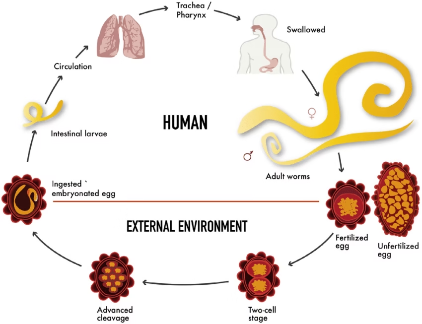
The life cycle of Ascaris lumbricoides is complex, involving several developmental stages both inside and outside the human body:
-
- Egg Stage:
- Ascaris eggs are passed in the feces of an infected person.
- The eggs become embryonated in the soil, requiring warmth and moisture to develop. It usually takes 2-4 weeks for the eggs to mature into infective larvae.
- Larval Stage:
- Humans become infected when ingest embryonated eggs from contaminated food, water, or hands.
- The eggs hatch in the small intestine, releasing larvae that burrow through the intestinal wall and enter the bloodstream.
- Migration:
- The larvae travel through the bloodstream to the lungs. In the lungs, they mature further and are carried up to the throat by the ciliary action of the bronchial tubes.
- The larvae are swallowed and enter the small intestine again, where they mature into adult worms.
- Adult Stage:
- In the small intestine, the larvae mature into adult worms.
- Female worms lay large eggs (up to 200,000 per day), passed in the feces.
- The life cycle continues when the eggs are released into the environment.
- Egg Stage:
Mode of Transmission
The transmission of Ascaris lumbricoides is primarily fecal-oral. Humans are infected by ingesting infective Ascaris eggs that are typically found on contaminated food, water, or hands. These eggs become embryonated in the environment and can survive in contaminated soil for months or even years.
Common modes of transmission include:
-
- Ingesting contaminated food or water.
- Hand-to-mouth transmission from contact with contaminated soil or surfaces.
- Poor hygiene practices, particularly in areas without proper sanitation or waste disposal systems.
Incubation Time
The incubation period for Ascaris lumbricoides varies depending on the stage of the infection:
-
- The eggs ingested by the host typically hatch in the small intestine within 6 hours to 2 days after ingestion.
- The larvae migrate through the lungs and back to the small intestine. This migration usually takes 1-2 weeks.
- Adult worms typically mature in the small intestine and begin laying eggs approximately 2-3 months after infection.
The full cycle from ingesting the egg to the laying of eggs by adult worms can take up to 2-3 months.
Pathogenesis
The clinical manifestations of ascariasis are often related to the migration of the larvae through the body and the presence of adult worms in the intestines. The pathology can vary based on the number of worms and the site of infection.
-
- Larval Migration:
- When larvae migrate through the lungs, they can cause Löffler’s syndrome, characterized by coughing, wheezing, and sometimes hemoptysis (coughing up blood). This is due to the irritation and inflammation of the lung tissue.
- Eosinophilia (elevated levels of eosinophils in the blood) is also commonly seen during the larval stage due to the body’s immune response.
- Adult Worms in the Intestine:
- As the worms mature in the small intestine, they can cause abdominal pain, nausea, vomiting, and diarrhea.
- Large numbers of worms can lead to intestinal blockage, particularly in children, and can also cause malabsorption of nutrients, leading to malnutrition.
- In severe cases, the worms can migrate to other body parts, such as the bile duct or appendix, causing further complications.
- Larval Migration:
Laboratory Diagnosis
The diagnosis of Ascaris lumbricoides infection is typically based on the identification of eggs or adult worms in stool samples or other diagnostic tests:
-
- Stool Examination:
- The most common method is the microscopic examination of stool samples for the presence of Ascaris eggs. The eggs are round, thick-walled, and have a characteristic bile-stained appearance.
- Eggs may take up to 2-3 months to appear in the stool after infection, so stool samples may need to be collected over several days or weeks.
- Serological Tests:
- Blood tests may show eosinophilia during the larval migration phase. However, eosinophilia is not specific to Ascaris and may be seen in other parasitic infections.
- Imaging Studies:
- In intestinal obstruction or migration of worms, imaging techniques such as X-rays, ultrasound, or CT scans can help locate the adult worms or visualize complications.
- Endoscopy:
- In rare cases, endoscopy may reveal adult worms in the intestine, though this is not a common diagnostic method.
- Stool Examination:
Treatment
The treatment of Ascaris lumbricoides infection primarily uses antihelminthic medications, effectively eliminating worms.
-
- Albendazole or Mebendazole:
- Both medications are effective against Ascaris and other intestinal nematodes. They work by inhibiting the absorption of glucose by the worms, leading to their eventual death.
- Albendazole is usually given as a single dose, while Mebendazole may require a 3-day course.
- Ivermectin:
- Ivermectin is an alternative treatment, particularly for individuals resistant to albendazole or mebendazole.
- Supportive Care:
- In severe cases, such as when there is an intestinal obstruction or other complications, surgical intervention or hospitalization may be required.
- Treatment may also involve addressing nutritional deficiencies if the infection has caused malabsorption or significant weight loss.
- Albendazole or Mebendazole:
Prevention
Preventing ascariasis relies on improving sanitation, hygiene, and access to clean water. Measures include:
-
- Proper sanitation and disposal of human waste.
- Ensuring clean water sources and avoiding the consumption of contaminated water.
- Wash hands thoroughly after using the toilet and before eating.
- Educating communities in endemic areas about proper hygiene practices and avoiding contaminated food.