Introduction
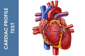
- A cardiac profile test is a comprehensive panel of blood tests designed to evaluate the heart’s overall health and detect any risk factors associated with cardiovascular diseases.
- It plays a crucial role in the early diagnosis, monitoring, and management of conditions such as coronary artery disease, myocardial infarction, and heart failure.
- Biomarkers such as lipid profile (total cholesterol, LDL, HDL, triglycerides), cardiac enzymes (like CK-MB, troponins), and inflammatory markers (such as C-reactive protein). Some advanced panels may also assess homocysteine levels, NT-proBNP, and lipoprotein (a), among others.
- By analyzing these parameters, clinicians can assess the extent of cardiac stress, inflammation, and the likelihood of atherosclerosis or ischemic events, enabling timely medical intervention and lifestyle modifications to prevent serious cardiac outcomes.
Cardiac Profile Test
-
Lipid Profile
-
Total Cholesterol – Measures the overall cholesterol in the blood.
-
LDL Cholesterol (Low-Density Lipoprotein) – “Bad” cholesterol contributing to plaque buildup in arteries.
-
HDL Cholesterol (High-Density Lipoprotein) – “Good” cholesterol that helps remove excess cholesterol from the blood.
-
Triglycerides – Type of fat (lipid) in the blood; elevated levels increase heart disease risk.
-
VLDL (Very Low-Density Lipoprotein) – Transports triglycerides; linked with atherosclerosis.
-
-
Cardiac Enzymes & Biomarkers
-
Troponin I or T – Highly specific and sensitive markers of myocardial injury (heart attack).
-
Creatine Kinase-MB (CK-MB) – Enzyme found in the heart; elevated in myocardial damage.
- Total CPK (Creatine Phosphokinase)
-
Myoglobin – Early marker of muscle injury, including cardiac muscle.
- SGOT – (AST – Aspartate Aminotransferase)
- LDH – (Lactate Dehydrogenase)
-
-
Inflammatory Marker
-
High-Sensitivity C-Reactive Protein (hs-CRP) – Indicates low-grade inflammation associated with cardiovascular risk.
-
-
Brain Natriuretic Peptide (BNP) or NT-proBNP
-
Used to assess and monitor heart failure severity.
-
-
Homocysteine
-
Elevated levels are linked to increased risk of coronary artery disease.
-
-
Lipoprotein (a)
-
Genetic lipid marker associated with early atherosclerosis and cardiovascular events.
-
-
Electrolytes and Minerals
-
Potassium, Sodium, Calcium, and Magnesium – Help evaluate cardiac function and rhythm disturbances.
-
Cardiac Enzymes & Biomarkers
Total Creatine Phosphokinase
Principle
Creatine Phosphokinase (CPK or CK) catalyzes the reversible transfer of a phosphate group from creatine phosphate to ADP, forming ATP and creatine:
Creatine phosphate+ADP→CPK→Creatine+ATP
ATP+Glucose→HK→Glucose-6-phosphate+ADP
The increase in NADPH concentration is directly proportional to CPK activity and is measured colourimetrically at 340 nm.
Method
Colourimetric
Type
Enzymatic assay
Sample Material
-
Specimen: Serum (preferred) or plasma (heparinized)
-
Collection: Avoid hemolysis (RBCs contain enzymes that interfere)
-
Storage: Analyze within a few hours; if delayed, refrigerate at 2–8°C (stable for 24 hours)
Normal Reference Range
-
Male: 38–174 U/L
-
Female: 26–140 U/L
Requirements
- Colorimeter or spectrophotometer (set at 340 nm)
- Water bath (37°C)
- Test Tube
- Stop watch
- Micropipettes
Reagents
R1
| Imidazole buffer, pH 6.1 | 125 mmol/l |
| Glucose | 25 mmol/l |
| Magnesium acetate | 12.5 mmol/l |
| EDTA | 2 mmol/l |
| N-acetylcysteine | 25 mmol/l |
| NADP | 2.4 mmol/l |
| Hexokinase | > 6.8 U/ml |
R2
| ADP | 15.2 mmol/l |
| D-glukoso-6-phosphate-dehydrogenase | > 8.8 U/ml |
| Creatine phosphate | 250 mmol/l |
| AMP | 25 mmol/l |
| Diadenosine pentaphosphate | 103 μmol/l |
Procedure
| Reagent 1 (buffer) | 1.000 ml |
| Sample | 0.050 ml |
Mix and incubate for 3 min. at 37°C. Then add:
| Reagent 2 (substrate) | 0.250 ml |
Mix and incubate for 3 min. at 37 °C. Then, measure the absorbance and, at the same time, start the stopwatch. Read the absorbance again exactly after 1, 2 and 3 minutes. Calculate the average 1-minute absorbance change (ΔA).
Monoreagent method – sample start
| Working solution | 1.000 ml |
| Sample | 0.040 ml |
Mix and incubate for 3 min. at 37 °C.
Calculation
CPK (U/L)=ΔA/min×F(F = factor provided by manufacturer)
Principle of Troponin Test
Troponins are regulatory proteins involved in calcium-mediated muscle contraction. The cardiac troponin complex has three subunits:
-
Troponin C (TnC) – binds calcium
-
Troponin I (TnI) – inhibits actin-myosin interaction
-
Troponin T (TnT) – binds the complex to tropomyosin
In cardiac muscle:
-
cTnI and cTnT have unique amino acid sequences, distinct from skeletal muscle isoforms.
-
During myocardial injury, especially acute myocardial infarction (AMI), cardiac cell membranes are disrupted, leading to leakage of intracellular contents, including troponins, into the circulation.
Detection:
-
The test is based on antibody-antigen interactions, where monoclonal or polyclonal antibodies specific to cTnI or cTnT are used.
-
The amount of troponin detected reflects the extent of myocardial injury.
Sample Type and Collection
-
Sample: Venous blood
-
Preferred tube: Serum (red-top) or plasma (lithium heparin or EDTA)
-
Storage:
-
Room temperature: up to 8 hours
-
Refrigerated (2–8°C): up to 24 hours
-
Frozen (−20°C or below): longer storage
-
Timing:
-
Troponins begin to rise: 3–6 hours after injury
-
Peak levels: 12–24 hours
-
Remain elevated: 7–10 days (cTnT may persist longer than cTnI)
Methods Used
A. Immunoassay-Based Methods (Routine clinical use)
-
Enzyme-Linked Immunosorbent Assay (ELISA)
-
Sandwich-type assay using capture and detection antibodies.
-
Signal is proportional to troponin concentration.
-
-
Chemiluminescent Immunoassay (CLIA)
-
Uses a chemiluminescent label that emits light during a reaction.
-
Higher sensitivity and automation.
-
-
Fluorescence Immunoassays (FIA)
-
Fluorescent tags emit signals read by detectors.
-
-
High-Sensitivity Troponin Assays (hs-cTn)
-
Can detect troponin levels in picogram/mL range
-
Allows early diagnosis (within 1–2 hours)
-
B. Point-of-Care Testing (POCT)
-
Rapid immunochromatographic tests
-
Useful in ER or ambulance settings
-
Less sensitive but faster (results in ~15 min)
C. Mass Spectrometry (Research Use)
-
High specificity and multiplexing ability
-
Rarely used in routine settings due to complexity
Clinical Significance:
-
Diagnosis of Myocardial Infarction (MI):
-
Troponin levels rise within 3–6 hours, peak at 12–24 hours, and may remain elevated for 7–10 days.
-
High-sensitivity assays can detect very early myocardial injury.
-
-
Differential Diagnosis of chest pain
-
Prognostic marker in:
-
Unstable angina
-
Heart failure
-
Pulmonary embolism
-
Sepsis-related cardiac injury
-
-
Risk stratification in cardiac patients
Creatine Kinase MB
Principle
Creatine phosphate + ADP —–CKBB / CKMB——> Creatine + ATP
ATP + Glucose——HK———-> Glucose-6-phosphate + ADP
Glucose-6-phosphate + NADP—–G6PDH—–> Gluconate-6-P + NADPH + H+
Method
Enzymatic method
Sample
Serum free of hemolysis, heparinized plasma, protected from light. Stability
24 hrs. at 2-8°C; 3 days at -20°C
Reagents
Reagent I: Buffer Reagent I
Reagent II: Enzyme Reagent II
Procedure
| Working Reagent | 1000 µl |
Incubate at Assay temperature for 1 min and add.
| Sample | 50 µl |
Mix well and after 5 min. measure the change in absorbance. Read the initial absorbance and start timer simultaneously, read again after every 60 seconds for 5 minutes. Calculate ∆ A/min.
Calculation
U/l CKB = ∆ A/min. x 6752
Lactate dehydrogenase
Principle
Kinetic determination of lactate dehydrogenase according to the following reaction.
Pyruvate + NADH + H+———-LDH———> L-Lactate +NAD+
Method
Optimized UV-test according to IFCC
Sample
- Serum or heparin plasma or EDTA plasma. Separate at the latest 1h after blood collection from cellular contents.
- 1 month at 4 – 8°C
- Discard contaminated specimens
Reagents
Reagent I: Buffer reagent
Reagent II: Substrate reagent
Procedure
| Working Reagent | 1000 μl |
| Sample | 25 μl |
Mix well and after 1 min incubation, measure the change in absorbance per min. (ΔA/min.) for next 2 minutes.
Calculation
LDH Activity (IU/L) = Δ A/min x 6592
SGOT (AST – Aspartate Aminotransferase)
CLICK HERE FOR MORE INFORMATION ——> SGOT