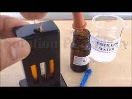
Various methods of estimation of Hb, by different methods, are a historical technique for estimating haemoglobin levels and are useful for quick assessments, especially in settings with limited resources. However, it is largely supplanted by more accurate and reliable methods in modern clinical practice.
Sahli’s method
Principle
Sahli’s method relies on the fact that haemoglobin in the blood can be converted into a coloured solution when red blood cells are lysed. The intensity of the colour produced correlates with the haemoglobin concentration in the sample.
Materials Required
- Blood Sample: Fresh venous blood (2-3 mL).
- Reagents:
- Hydrochloric Acid (HCl): Concentration of about 0.1 N.
- Distilled Water: For dilution and washing.
- Glassware:
- Test tubes (at least two for the sample and one for the standard).
- Pipettes (for accurate measurement of liquids).
- A graduated cylinder (optional for larger measurements).
- Hemoglobinometer or Color Comparison Chart: A device or printed chart to compare the colour of the hemolysate against known standards.
Procedure
Step 1: Preparation of Blood Sample
- Collect the Blood:
- Collect venous blood into a clean test tube or container using an aseptic technique.
- If using an anticoagulant (like EDTA), ensure the blood remains fluid for accurate measurement.
Step 2: Hemolysis of Red Blood Cells
- Add Hydrochloric Acid:
- Measure about 2 mL of blood and transfer it to a test tube.
- Carefully add 2-3 drops of 0.1 N hydrochloric acid to the blood sample. This acid will lyse the red blood cells, releasing haemoglobin into the solution.
- Mix the Solution:
- Gently invert the test tube or mix the blood and acid solution with a pipette.
- Allow the mixture to stand at room temperature for 5-10 minutes. The solution will turn red as the haemoglobin is released and dissolved.
Step 3: Preparation of the Standard
- Prepare a Standard Solution:
- If a standard solution is unavailable, prepare a standard haemoglobin solution with a known concentration (e.g., 15 g/dL).
- This can be done by dissolving a specific amount of purified haemoglobin in distilled water to create a colour standard for comparison.
Step 4: Color Comparison
- Dilution (if necessary):
- If the hemolysate is too concentrated, dilute it with distilled water to achieve a suitable concentration for comparison. The typical dilution ratio is about 1:10, but this can be adjusted based on the depth of colour.
- Use a Second Test Tube:
- Transfer an equal volume (2 mL) of the standard haemoglobin solution into a separate test tube for comparison.
- Comparison of Colors:
- Place both test tubes (the hemolysate and the standard solution) against a white background or light source to enhance visibility.
- Compare the colour of the hemolysate with the standard solution or colour comparison chart. Look for the closest match in colour intensity.
Step 5: Interpretation of Results
- Record the Hemoglobin Level:
- Based on the comparison, estimate the haemoglobin concentration in the blood sample. The concentration is typically expressed in grams per deciliter (g/dL).
- Use the colour intensity of the hemolysate to determine the haemoglobin level by referencing the standard.
Advantages
- Simple and Cost-Effective: Requires minimal equipment and can be performed in basic laboratory settings.
- Quick Procedure: Results can be obtained relatively quickly.
Disadvantages
- Subjective Measurement: Color comparison can be affected by lighting conditions and personal interpretation, leading to potential inaccuracies.
- Limited Precision: Less accurate than advanced methods, especially in hemoglobinopathies or abnormal haemoglobin forms.
- Interferences: Other blood components can influence the colour, impacting the accuracy of the results.
Principle of the Cyanmethemoglobin Method
The Cyanmethemoglobin method is based on converting haemoglobin (Hb) in the blood to a stable, coloured complex known as cyanmethemoglobin. This conversion occurs through the action of specific reagents, primarily potassium ferricyanide and potassium cyanide.
Materials Required
- Blood Sample: Fresh venous blood (5-10 mL).
- Reagents:
- Drabkin’s Solution: A mixture containing:
- Potassium ferricyanide (K₃[Fe(CN)₆]): Converts haemoglobin to cyanmethemoglobin.
- Potassium cyanide (KCN): Stabilizes the cyanmethemoglobin complex.
- Distilled Water: For dilution and washing.
- Drabkin’s Solution: A mixture containing:
- Glassware:
- Test tubes.
- Pipettes (for accurate measurement of liquids).
- Cuvettes (for spectrophotometric measurement).
- Spectrophotometer: For measuring the absorbance at a specific wavelength (540 nm).
- Standard Hemoglobin Solution: For calibration and comparison.
Procedure
Step 1: Preparation of Blood Sample
- Collect the Blood:
- Use an aseptic technique to collect venous blood into a clean test tube containing an anticoagulant (like EDTA) to prevent clotting.
Step 2: Preparation of Hemolysate
- Add Drabkin’s Solution:
- Measure 5 mL of Drabkin’s solution and add it to a test tube.
- Add 20 µL (0.02 mL) of the blood sample to the Drabkin solution test tube.
- Mix the Solution:
- Mix the solution by inverting the test tube or using a vortex mixer to ensure thorough mixing. This process lyses the red blood cells and converts haemoglobin to cyanmethemoglobin.
- Allow Reaction to Occur:
- Let the mixture stand for 10-15 minutes at room temperature. This allows for the complete conversion of haemoglobin to cyanmethemoglobin.
Step 3: Measurement
- Set Up the Spectrophotometer:
- Turn on the spectrophotometer and allow it to warm up if necessary.
- Calibrate the spectrophotometer using distilled water as a blank. Set the wavelength to 540 nm.
- Measure the Absorbance:
- Transfer the prepared solution to a cuvette.
- Place the cuvette in the spectrophotometer and record the absorbance at 540 nm.
Step 4: Calculation
- Standard Curve:
- Prepare a standard curve using known concentrations of haemoglobin. Measure their absorbance at 540 nm and plot the results to create a standard curve.
- Calculate Hemoglobin Concentration:
- Use the absorbance value from the test sample and compare it to the standard curve to determine the haemoglobin concentration in grams per deciliter (g/dL).
Advantages
- High Accuracy: The method provides precise and reliable measurements of haemoglobin levels.
- Wide Applicability: Can differentiate between various haemoglobin types with additional testing.
- Standardization: Well-established method with standardized protocols.
Disadvantages
- Handling of Cyanide: Potassium cyanide is toxic, requiring careful handling and disposal.
- Time-Consuming: Takes longer than some point-of-care tests, requiring 15-20 minutes to complete.
- Interferences: Certain substances (e.g., elevated bilirubin or lipemia) can affect absorbance measurements.
Errors Involved in Hemoglobin Estimation
1. Pre-Analytical Errors
- Sample Collection:
-
- Aseptic Technique: Not using an aseptic technique can lead to contamination. Blood samples should be collected using sterile equipment and proper skin antisepsis.
- Vascular Complications: Incorrect venipuncture can result in hemolysis. Use a gentle approach to avoid damaging red blood cells.
- Storage Conditions:
-
- Time Delay: If blood samples are not processed promptly, haemoglobin can degrade, leading to inaccurate results. It is recommended to analyze samples within 1-2 hours of collection.
- Temperature: If processing is delayed, samples should be kept at 2-8°C. Prolonged exposure to room temperature can cause hemolysis.
- Anticoagulant Choice:
-
- EDTA vs. Citrate: An inappropriate anticoagulant can alter the sample’s haemoglobin behaviour. EDTA is commonly used for haematological tests.
2. Analytical Errors
- Reagent Preparation:
-
- Concentration Issues: Incorrect preparation of Drabkin’s solution can lead to inaccurate results. Regular checks on the reagent concentration are vital.
- Expiration: Using expired reagents can result in ineffective hemolysis or the formation of the cyanmethemoglobin complex.
- Inadequate Mixing:
-
- Incomplete Hemolysis: If the blood sample is not thoroughly mixed with the reagent, the hemolysis may be incomplete, affecting the accuracy of the measurement.
- Spectrophotometer Calibration:
-
- Wavelength Accuracy: Ensure that the spectrophotometer is calibrated to the correct wavelength (540 nm) for optimal measurement of cyanmethemoglobin.
- Absorbance Range: Regularly check the instrument’s absorbance range to ensure it functions within the specified limits.
- Interference from Other Substances:
-
- Bilirubin and Lipemia: High levels of bilirubin or lipids can interfere with the colourimetric measurement, leading to falsely elevated or decreased haemoglobin readings. Pre-treatment of samples may be necessary in such cases.
3. Post-Analytical Errors
- Data Interpretation:
-
- Reference Ranges: Results can be misinterpreted if the laboratory does not use appropriate reference ranges based on the tested population.
- Variability: Variability in results may arise from differences in methodology or equipment between laboratories.
- Human Error:
-
- Pipetting Errors: Inaccurate pipetting can lead to errors in sample and reagent volumes. Ensure proper techniques and use calibrated pipettes.
- Calculation Mistakes: Errors in calculating haemoglobin concentration from absorbance values can skew results.
Standardization of Instruments for Hemoglobin Estimation
1. Calibration of Spectrophotometer
- Blank Calibration:
-
- Use distilled water or a blank of the reagent solution (Drabkin’s solution) to set the baseline absorbance to zero. This step ensures that only the absorbance from the haemoglobin is measured.
- Standard Curve Preparation:
-
- Create a standard curve using several known haemoglobin concentrations (e.g., 0, 5, 10, 15 g/dL). Measure the absorbance of these standards to establish a relationship between absorbance and concentration.
- Plot the absorbance against haemoglobin concentration to derive the equation for future calculations.
2. Quality Control
- Control Samples:
-
- Use quality control samples with known haemoglobin concentrations alongside patient samples. Regular testing of controls helps monitor the accuracy and precision of the method.
- Analyze control samples daily to ensure the system is functioning correctly.
- Documentation:
-
- Maintain a log of all control measurements, calibration data, and any adjustments made to the equipment or procedures. This documentation is crucial for tracking performance over time.
3. Routine Maintenance
- Cleaning:
-
- Regularly clean cuvettes and the spectrophotometer to prevent contamination and ensure consistent results. Follow the manufacturer’s recommendations for cleaning procedures.
- Professional Servicing:
-
- Schedule periodic servicing and calibrating of the spectrophotometer by qualified technicians to maintain accuracy and reliability.
4. Training of Personnel
- Standard Operating Procedures (SOPs):
-
- Develop and implement clear SOPs for all aspects of haemoglobin testing, including specimen collection, reagent preparation, measurement, and interpretation.
- Ensure that all laboratory personnel know and adhere to these SOPs.
- Training Programs:
-
- Provide regular training for staff on the latest techniques, equipment use, and error-reduction strategies. Continuous education helps reduce human error and improve overall laboratory performance.