Introduction
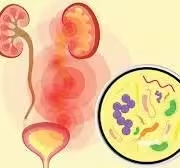
- Urinary tract infections (UTIs) are common infections that can affect any part of the urinary system, including the bladder (cystitis) and kidneys (pyelonephritis).
- UTIs can be caused by various bacteria, with Escherichia coli being the most prevalent.
- Accurate laboratory diagnosis is essential for effective treatment and management.
Clinical Presentation
Common Symptoms
-
Lower UTI (Cystitis):
- Dysuria: Painful urination is often described as a burning sensation.
- Urgency: A strong, persistent need to urinate.
- Frequency: Increased need to urinate, often producing small volumes.
- Suprapubic Pain: Discomfort in the lower abdomen.
- Hematuria: Blood in urine, which may be visible or detected microscopically.
-
Upper UTI (Pyelonephritis):
- Flank Pain: Pain in the back and side, often severe.
- Fever and Chills: Indicative of a systemic response to infection.
- Nausea and Vomiting: Associated gastrointestinal symptoms may occur.
- Malaise: General feelings of unwellness.
Sample Collection
-
Urine Sample Types
- Mid-Stream Clean Catch Urine: The preferred method for outpatient settings to minimize contamination.
- Catheterized Urine: Useful for patients who cannot provide a clean catch sample or in cases of suspected catheter-associated UTI.
- Suprapubic Aspiration: A sterile procedure for obtaining urine directly from the bladder, often used in infants or research settings.
-
Collection Technique
- Proper technique is critical:
- Preparation: Instruct the patient to wash their hands and clean the genital area.
- Collection: Instruct the patient to start urinating, then collect the mid-stream portion of urine in a sterile container.
- Minimizing Contamination: Avoid contact with the skin or external genitalia.
-
Transport and Handling
- Urine samples should ideally be processed within 2 hours of collection. If this is not possible:
- Refrigeration: Store samples at 4°C if processing is delayed.
- Preservatives: Boric acid can be added to urine samples to stabilize bacterial counts.
Laboratory Techniques
Urinalysis
A. Physical Examination
- Color: Normal urine is pale yellow; darker urine can indicate dehydration, while cloudy urine suggests infection or crystals.
- Clarity: Urine should be clear; turbidity may suggest infection or the presence of crystals.
- Odor: Foul-smelling urine may indicate infection.
B. Chemical Examination
- Dipstick Testing: A rapid method to screen for abnormalities:
- Leukocyte Esterase: Indicates white blood cells; a positive result suggests infection.
- Nitrites: The presence of nitrites indicates the conversion of nitrates by bacteria, commonly seen in E. coli infections.
- Blood: This may indicate injury, stones, or infection.
- Protein: This may be present in infections or kidney disease.
C. Microscopic Examination
- A spun urine sample is analyzed:
- White Blood Cells (WBCs): Counts >5 WBCs per high power field (HPF) are suggestive of infection.
- Red Blood Cells (RBCs) may indicate glomerular disease or injury.
- Bacteria: The presence of bacteria confirms suspicion of infection.
- Casts: May indicate kidney involvement, such as in pyelonephritis.
Urine Culture
A. Culture Procedure
- Media Selection:
- MacConkey Agar: Selective for Gram-negative bacteria; lactose fermenters turn pink.
- CLED Agar: Supports growth of uropathogens and inhibits swarming of Proteus species.
B. Incubation Conditions
- Incubate cultures at 35-37°C for 24-48 hours. Aerobic conditions are essential, and some media may also be incubated in CO₂ for certain organisms.
C. Identification of Organisms
- Colony Count: A significant growth is defined as >10^5 CFU/mL for a UTI diagnosis.
- Colony Morphology: Color, shape, and size help identify the type of bacteria.
- Biochemical Tests: Further testing can include:
- Oxidase Test: Differentiates between oxidase-positive organisms (e.g., Pseudomonas) and others.
- Urease Test: Identifies Proteus species that produce urease.
- Indole Test: Identifies E. coli based on indole production from tryptophan.
Antimicrobial Susceptibility Testing
- Essential for guiding treatment, particularly in complicated UTIs or recurrent infections:
- Disk Diffusion Method (Kirby-Bauer): Involves placing antibiotic disks on an agar plate inoculated with the organism; the zone of inhibition indicates susceptibility.
- Broth Microdilution: Provides minimum inhibitory concentration (MIC) values, which help determine the lowest concentration of antibiotic that prevents growth.
Interpretation of Results
Urinalysis
- Positive Dipstick Tests: The presence of leukocyte esterase and nitrites strongly indicates a UTI.
- Microscopic Findings: High numbers of WBCs, bacteria, and potential casts suggest a urinary tract infection.
Urine Culture
- Positive Culture: Isolation of a pathogen in significant quantities (>10^5 CFU/mL).
- Common Pathogens:
- Escherichia coli: The predominant cause (up to 80-90% of cases).
- Klebsiella pneumoniae: Common in catheter-associated UTIs.
- Proteus mirabilis: Notable for urease production and associated with alkaline urine.
- Enterococcus species May occur in complicated cases or in hospitalized patients.
- Pseudomonas aeruginosa: Often seen in complicated UTIs or among patients with catheter use.
Antibiotic Sensitivity
- Antibiotic susceptibility profiles guide empirical therapy, particularly in cases of multidrug-resistant organisms or recurrent infections.
Clinical Implications
Treatment
- Early diagnosis allows for prompt initiation of antibiotics. Common empirical treatments include:
- Nitrofurantoin: Effective for uncomplicated cystitis.
- Trimethoprim-sulfamethoxazole (TMP-SMX): Effective for many UTI pathogens, but resistance is increasing.
- Fosfomycin: Useful for uncomplicated cases and has a single-dose regimen.
- For pyelonephritis or complicated UTIs, broader-spectrum antibiotics may be required:
- Ciprofloxacin or Levofloxacin: Fluoroquinolones used for more severe infections.
- Ceftriaxone: Often used for IV therapy in hospitalized patients.
Follow-Up
- In patients with recurrent UTIs, further evaluation may be warranted. This can include:
- Imaging Studies: Ultrasound or CT scans to check for anatomical abnormalities, kidney stones, or obstructions.
- Urology Consultation: In cases of recurrent infections or complications, referral for further evaluation and potential intervention may be necessary.
Prevention Strategies
- Education on hygiene practices (e.g., wiping front to back, urinating after intercourse).
- Encouraging increased fluid intake to promote regular urination.
- Prophylactic antibiotics may sometimes be prescribed for individuals with recurrent UTIs.