Introduction
- Trypanosomes are microscopic, flagellated protozoan parasites belonging to the genus Trypanosoma.
- They are important blood and tissue parasites that infect humans and various animals.
- The two major human pathogenic species are Trypanosoma brucei and Trypanosoma cruzi.
- T. brucei causes African trypanosomiasis (sleeping sickness) and is transmitted mainly by the tsetse fly.
- T. cruzi causes Chagas disease (American trypanosomiasis) and is mainly transmitted by triatomine or kissing bugs.
- Trypanosomes have characteristic morphological features such as a kinetoplast, flagellum, and undulating membrane.
- Diagnosis may involve microscopic examination, concentration techniques, serological tests, and molecular methods, depending on the species and stage of infection.
Habitat
Trypanosomes are extracellular parasites that typically reside in the bloodstream or tissues of their vertebrate hosts. The habitat of the parasite depends on the species and the disease it causes:
- African Trypanosomes (Trypanosoma brucei): Found in the blood of humans and animals in sub-Saharan Africa. The disease is transmitted by the tsetse fly (genus Glossina).
- American Trypanosomes (Trypanosoma cruzi): Found in the blood of humans and animals in Central and South America. It is transmitted by triatomine bugs (known as kissing bugs, genus Triatoma).
Trypanosomes also live in the insect vectors, undergoing certain developmental stages, including multiplication and maturation.
Epidemiology
The epidemiology of trypanosomiasis is largely dependent on the region of the world and the specific type of trypanosome.
- African Trypanosomiasis (Sleeping Sickness):
- Caused by Trypanosoma brucei species (specifically T. b. gambiense and T. b. rhodesiense).
- Endemic areas: Primarily sub-Saharan Africa, with some outbreaks in regions of Central Africa and East Africa.
- Vectors: Transmitted by the tsetse fly, which lives in rural, forested, or savanna areas.
- Human-to-human transmission is possible, particularly with T. b. gambiense through the bite of an infected fly or from mother to child during pregnancy or childbirth.
- American Trypanosomiasis (Chagas Disease):
- Caused by Trypanosoma cruzi.
- Endemic areas: Found in rural areas of Latin America, including countries like Brazil, Argentina, Mexico, and Bolivia.
- Vectors: Transmitted by triatomine bugs (kissing bugs) found in cracks and crevices of homes.
- Transmission can also occur via blood transfusion, organ transplantation, or from mother to child during pregnancy.
Morphology
Trypanosomes exhibit characteristic morphological forms during their life cycle. These forms are based on the location within the host or vector:
- Trypomastigote (blood stage in mammals and tsetse fly stage):
- This is the infective form that circulates in the bloodstream of mammals.
- It has a long, slender body, a single flagellum that extends along the body, and a prominent kinetoplast (a DNA-containing structure in the mitochondrion).
- The flagellum is located at the anterior end, extending as a free flagellum or attached along the body.
- Epimastigote (intermediate stage in the vector):
- This form is found in the midgut of the tsetse fly or triatomine bug.
- It has a shorter body with a flagellum extending from the anterior end.
- The kinetoplast is located near the anterior region of the cell.
- Amastigote (tissue stage in mammals):
- Found in tissues of the mammalian host (e.g., heart, liver, spleen).
- It is oval-shaped and non-flagellated.
- Amastigotes multiply by binary fission in tissue macrophages.
Life Cycle
The life cycle of trypanosomes is complex and involves two hosts: a vertebrate host (usually a mammal) and an insect vector (either the tsetse fly for African trypanosomes or the triatomine bug for American trypanosomes). The life cycle consists of several stages, including transformation from one form to another.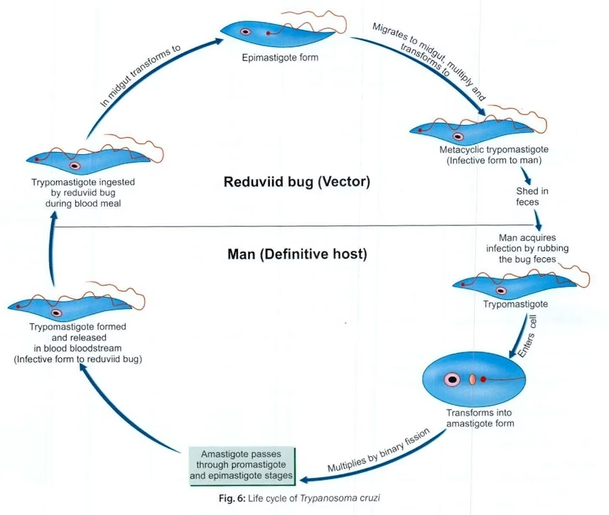
- In the Mammalian Host:
- The trypomastigote is injected into the bloodstream when an infected insect vector bites the host.
- The trypomastigotes circulate in the bloodstream and invade various tissues, including the heart, liver, spleen, and lymph nodes.
- Sometimes, the trypomastigotes transform into amastigotes within the host’s tissues, multiplying by binary fission.
- The amastigotes later differentiate into trypomastigotes, which re-enter the bloodstream.
- In the Insect Vector:
- The vector (tsetse fly or triatomine bug) becomes infected by ingesting trypomastigotes during a blood meal.
- The trypomastigotes transform into epimastigotes in the insect’s gut.
- The epimastigotes multiply and later transform into trypomastigotes.
- These infective trypomastigotes migrate to the salivary glands or the hindgut of the insect, ready to be transmitted back to a mammalian host during the next bite.
Pathogenesis
African Trypanosomiasis (Sleeping Sickness):
-
- T. b. gambiense causes the chronic form of the disease, which progresses slowly over several years, while T. b. rhodesiense causes the acute form, leading to more rapid progression.
- The parasite is initially localized in the bloodstream, causing fever, headaches, and lymphadenopathy (swollen lymph nodes).
- As the disease progresses, neurological symptoms appear, such as drowsiness, confusion, and coma, due to the invasion of the central nervous system (CNS). The patient eventually falls into a coma, hence the name sleeping sickness.
American Trypanosomiasis (Chagas Disease):
-
- The parasite initially infects the heart, gut, and nervous system.
- In the acute phase, symptoms include fever, swelling at the bite site (chagoma), and lymphadenopathy.
- In the chronic phase, the infection can lead to severe complications such as heart failure, dilated cardiomyopathy, megaesophagus, and megacolon.
The parasites evade the immune system through antigenic variation, meaning they periodically change the surface proteins recognized by the host immune system.
Laboratory Diagnosis
Laboratory diagnosis of trypanosomiasis is based on demonstration of the parasite, detection of antibodies, or detection of parasite DNA. The choice of test depends on the species, stage of infection, and clinical presentation.
1. Specimens Used
The clinical specimen depends on the suspected infection:
- Peripheral blood – commonly used for detection of bloodstream trypomastigotes.
- Lymph node aspirate – useful in African trypanosomiasis, especially when lymphadenopathy is present.
- Chancre fluid – may be examined during early African trypanosomiasis.
- Cerebrospinal fluid (CSF) – examined when neurological involvement is suspected.
- Serum – mainly used for serological diagnosis of T. cruzi infection.
2. Direct Microscopic Examination
Microscopy is an important method for detecting trypomastigotes.
A. Wet Mount
- Fresh blood or other appropriate specimens are examined under the microscope.
- Live trypanosomes may be recognized by their characteristic active motility.
- The method is simple but may have limited sensitivity when parasitemia is low.
B. Thick Blood Film
- A relatively large volume of blood is examined.
- It is useful for detecting parasites when their number is low.
- Once detected, further examination can help establish the morphology.
C. Thin Blood Film
A thin blood smear allows better observation of parasite morphology.
Important features include:
- Elongated body
- Nucleus
- Kinetoplast
- Undulating membrane
- Flagellum
3. Concentration Methods
When the number of parasites in blood is low, concentration techniques can improve detection.
Common methods include:
- Microhematocrit centrifugation
- Buffy-coat examination
- Other validated parasite concentration techniques
These methods are particularly useful in the diagnosis of African trypanosomiasis.
4. Lymph Node Aspirate Examination
- Lymph node aspiration can be performed when lymphadenopathy is present.
- The aspirated material is examined microscopically.
- Motile trypanosomes may be demonstrated.
- This method can be particularly useful in gambiense African trypanosomiasis.
5. Examination of Cerebrospinal Fluid
CSF examination is important when neurological involvement is suspected in African trypanosomiasis.
The examination may include:
- Detection of trypanosomes
- White blood cell count
- Other CSF parameters used for assessment of neurological-stage disease
CSF findings are interpreted together with clinical findings and other diagnostic tests.
6. Serological Diagnosis
Serological tests detect antibodies against Trypanosoma.
They are particularly important for Chagas disease caused by T. cruzi.
Common approaches include:
- Enzyme immunoassays
- Indirect immunofluorescence assays
- Other validated antibody-based tests
Important: In chronic Chagas disease, diagnosis generally relies on serological testing because parasite levels in peripheral blood may be very low.
7. Molecular Diagnosis
Polymerase chain reaction (PCR) can detect Trypanosoma DNA in appropriate clinical specimens.
Molecular methods may be useful for:
- Detection of infection
- Species identification
- Detection of low-level parasitemia
- Selected cases where conventional methods are inconclusive
PCR availability and clinical utility vary according to the laboratory and clinical setting.
8. Xenodiagnosis
- Xenodiagnosis is a specialized method historically used mainly for Chagas disease.
- Laboratory-reared, uninfected triatomine bugs are allowed to feed on the patient.
- The insects are subsequently examined for T. cruzi.
- It is now largely restricted to specialized or research settings because more practical diagnostic methods are available.
Treatment
Treatment of trypanosomiasis depends on the species of Trypanosoma, stage of infection, and presence of neurological involvement.
1. Treatment of African Trypanosomiasis
- African trypanosomiasis is caused by Trypanosoma brucei.
- A. T. b. gambiense infection
Depending on disease stage and current treatment recommendations, drugs may include:
- Fexinidazole – used for appropriate cases, including selected patients with neurological-stage disease.
- Pentamidine – commonly used for early-stage disease.
- Eflornithine + Nifurtimox (NECT) – an important treatment option for appropriate neurological-stage disease.
B. T. b. rhodesiense infection
Treatment options include:
- Suramin – mainly used for early-stage disease.
- Melarsoprol – used for neurological-stage disease.
Treatment should be selected according to the infecting subspecies and disease stage because some drugs do not adequately penetrate the central nervous system.
2. Treatment of Chagas Disease
- Chagas disease is caused by Trypanosoma cruzi.
The two principal antiparasitic drugs are:
A. Benznidazole
- One of the main drugs used to treat T. cruzi infection.
- Particularly effective when treatment is started early.
- Treatment duration is usually several weeks.
- Adverse effects may include skin reactions and peripheral neuropathy.
B. Nifurtimox
- Another important antiparasitic drug used against T. cruzi.
- Treatment is generally given for an extended period.
- Gastrointestinal and neurological adverse effects may occur.
Important: Treatment decisions should consider age, pregnancy, stage of infection, comorbidities, and potential drug toxicity.
Prevention
There is currently no widely available vaccine for human trypanosomiasis. Prevention therefore focuses mainly on vector control, screening, and reducing exposure.
1. Vector Control
Reducing contact with insect vectors is an important preventive measure.
For African Trypanosomiasis
- Control tsetse fly populations in endemic areas.
- Use insect traps and other vector-control measures.
- Avoid areas with high tsetse-fly activity when possible.
For Chagas Disease
- Control triatomine bugs using appropriate insecticides.
- Improve housing conditions to reduce insect hiding places.
- Repair cracks in walls and roofs where vectors may live.
2. Personal Protection
People living in or travelling to endemic areas should:
- Wear long-sleeved clothing and long trousers.
- Use appropriate insect repellents.
- Avoid unnecessary exposure to vector-infested areas.
- Use suitable insect barriers where appropriate.
3. Blood Donor Screening
Screening of blood donors is important to prevent transfusion-associated transmission, particularly for T. cruzi in areas where Chagas disease occurs.
4. Organ Donor Screening
Appropriate screening of organ donors can reduce the risk of transplant-associated transmission of T. cruzi.
5. Prevention of Congenital Transmission
- Women at risk of T. cruzi infection can be appropriately screened.
- Infants born to infected mothers should receive appropriate testing and follow-up.
- Confirmed congenital infection can be treated early, when treatment is generally more effective.
6. Food and Environmental Hygiene
For Chagas disease, transmission can occasionally occur through contaminated food or beverages.
Preventive measures include:
- Maintaining good food hygiene.
- Protecting food from insect contamination.
- Using safe drinking water.
- Following appropriate food preparation practices in endemic areas.
7. Early Detection and Treatment
Early diagnosis and treatment help:
- Reduce complications.
- Prevent progression of disease.
- Reduce the duration of parasitemia.
- Help prevent transmission in certain settings