- Filariae are a group of parasitic nematodes (roundworms) responsible for various human diseases known as filarial infections.
- The diseases caused by filariae are generally transmitted through the bites of infected arthropod vectors.
- They can lead to debilitating conditions such as lymphatic filariasis, onchocerciasis, and loiasis.
- The filariae’s life cycle involves a vertebrate host and an arthropod vector.
Habitat
Filarial worms are extracellular parasites in their vertebrate hosts’ tissues or lymphatic systems. The specific habitat varies depending on the species:
- Lymphatic filariasis (Wuchereria bancrofti, Brugia malayi, Brugia timori): Found in the host’s lymphatic vessels and lymph nodes.
- Onchocerciasis (Onchocerca volvulus): Found in subcutaneous tissues of the host, particularly around the eyes and in the skin.
- Loiasis (Loa loa): Found in subcutaneous tissues, especially in the eye and muscle tissue.
- Mansonella species: Found in subcutaneous tissues and body cavities.
The larvae or microfilariae are present in the blood or tissues of their vector hosts (arthropods like mosquitoes, black flies, and deerflies).
Epidemiology
Several factors, including geographic location, climate, and the presence of appropriate arthropod vectors, influence the epidemiology of filarial diseases.
- Lymphatic Filariasis:
- Caused by: Wuchereria bancrofti, Brugia malayi, Brugia timori.
- Endemic regions: Found in tropical and subtropical regions of Africa, Asia, Pacific Islands, and Central America.
- Vectors: Mosquitoes (e.g., Culex, Aedes, Anopheles).
- Transmission: Transmission occurs when an infected mosquito takes a blood meal from a human, injecting infective larvae (L3) into the bloodstream.
- Onchocerciasis (River Blindness):
- Caused by: Onchocerca volvulus.
- Endemic regions: Found in sub-Saharan Africa, Central America, and South America.
- Vectors: Blackflies (Simulium species) that breed in fast-moving streams and rivers.
- Transmission: The larvae (microfilariae) are transmitted to humans through the bite of an infected blackfly.
- Loiasis:
- Caused by: Loa loa.
- Endemic regions: Found in West and Central Africa.
- Vectors: Deerflies (Chrysops species).
- Transmission: Infected deerflies transmit infective larvae when they bite humans.
- Mansonellosis:
- Caused by: Mansonella ozzardi, Mansonella perstans, Mansonella streptocerca.
- Endemic regions: Found in tropical regions of Africa, South America, and Central America.
- Vectors: Midges or mosquitoes.
Morphology
Filarial nematodes have a characteristic elongated, cylindrical body and exhibit various forms during their life cycle:
- Adult worms (macrofilariae):
- Long and thread-like, ranging from 2 mm to 10 cm long, depending on the species.
- Male worms are usually smaller than females and may have a characteristic curved posterior end.
- Female worms are often longer and can produce large numbers of microfilariae.
- Microfilariae:
- The larval form found in the host’s blood or tissues is non-infective.
- They are thread-like and vary in size, with some having sheaths and others not.
- In the bloodstream, microfilariae move in a characteristic rhythmic manner.
- Infective larvae (L3):
- The infective larval stage is introduced into the human host through the bite of an infected vector.
- These larvae can enter the lymphatic system or subcutaneous tissues depending on the species.
Life Cycle
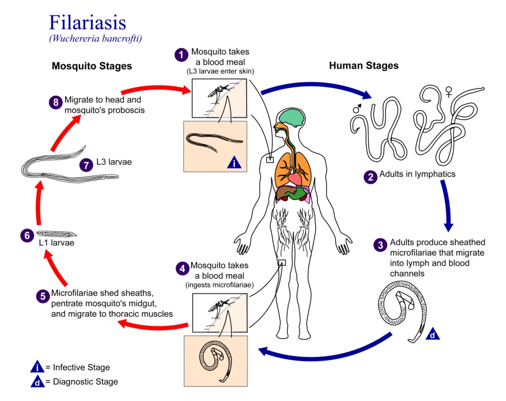
The life cycle of filarial worms is typically complex, involving a vertebrate host (such as humans) and an arthropod vector (mosquitoes, blackflies, or deerflies).
- In the Vertebrate Host:
- Infective larvae (L3) are injected into the host when the vector (e.g., mosquito, blackfly) takes a blood meal.
- The larvae migrate through the bloodstream and settle in specific tissues:
- Lymphatic system (in the case of Wuchereria and Brugia species).
- Subcutaneous tissue (in the case of Onchocerca and Loa species).
- The larvae develop into adult worms in the tissues, with females producing microfilariae (immature larvae).
- Depending on the species, these microfilariae can migrate to different tissues, including the bloodstream or skin.
- In the Arthropod Vector:
- An arthropod vector ingests the microfilariae during a blood meal.
- Inside the vector, the microfilariae develop into larval forms (L1, L2, and eventually L3, which is the infective form).
- The L3 larvae migrate to the vector’s mouthparts and are ready to infect another vertebrate host when the vector bites.
Pathogenesis
The pathogenesis of filarial infections varies depending on the species and the organs involved. Filarial diseases are typically characterized by chronic inflammation and immune response:
- Lymphatic Filariasis:
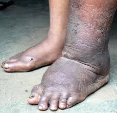
- The adult worms obstruct the lymphatic vessels, leading to lymphoedema and elephantiasis (swelling of limbs).
- Chronic inflammation can result in fibrosis and scarring of lymphatic vessels.
- Acute episodes of inflammation (e.g., acute filarial lymphangitis) occur due to microfilariae or the release of endotoxins from dying worms.
- Onchocerciasis:
- The presence of adult worms in the subcutaneous tissues causes granulomatous inflammation and the formation of nodules.
- The microfilariae migrate through the skin and eyes, leading to dermatitis, pruritus (itching), and in severe cases, blindness due to corneal opacification (river blindness).
- Intraocular inflammation can lead to retinal damage and optic nerve degeneration.
- Loiasis:
- Loa loa worms migrate through the subcutaneous tissues, often leading to eye involvement where worms may be seen in the conjunctiva (a condition known as Calabar swellings).
- It can cause subcutaneous swelling, itching, and, in rare cases, encephalopathy due to the migration of worms through the central nervous system.
- Mansonellosis:
- This is often a subclinical infection but can cause mild symptoms such as pruritus, rashes, and lymphadenopathy.
- Rarely, it may cause complications such as abdominal pain or joint pain.
Laboratory Diagnosis
Diagnosis of filarial infections is based on detecting microfilariae in blood or tissue samples and may involve serological, molecular, and microscopic techniques.
Microscopy:
- Blood Smears:
- Thick and thin blood smears detect microfilariae in the blood. The smears are stained with Giemsa or Wright’s stain.
- Night-time blood collection may be required for certain species, such as Wuchereria bancrofti, which has nocturnal periodicity.
- Onchocerca volvulus can be diagnosed by finding microfilariae in skin snips.
Serology:
- ELISA and Western blot tests can detect antibodies or antigens specific to filarial species.
- Immunochromatographic tests are also available for diagnosing lymphatic filariasis.
PCR:
- Polymerase Chain Reaction (PCR) is increasingly used for the detection and species identification of filarial worms, particularly in subclinical cases or where microfilariae are not readily detected.
Imaging:
- Ultrasonography is useful for detecting adult filarial worms in tissues like the scrotum (in the case of Wuchereria bancrofti) or nodules in onchocerciasis.