Introduction
- The liver is the largest internal organ and a vital gland in the human body. It performs over 500 essential functions, including bile production, metabolism, detoxification, and nutrient storage.
- Structurally, it is a highly vascular organ with specialized cells and units that allow it to carry out these functions efficiently.
External Structure of the Liver
The liver is a reddish-brown, wedge-shaped organ in the abdomen’s right upper quadrant (RUQ), just below the diaphragm. It is divided into lobes, surfaces, and ligaments that support and anchor it.
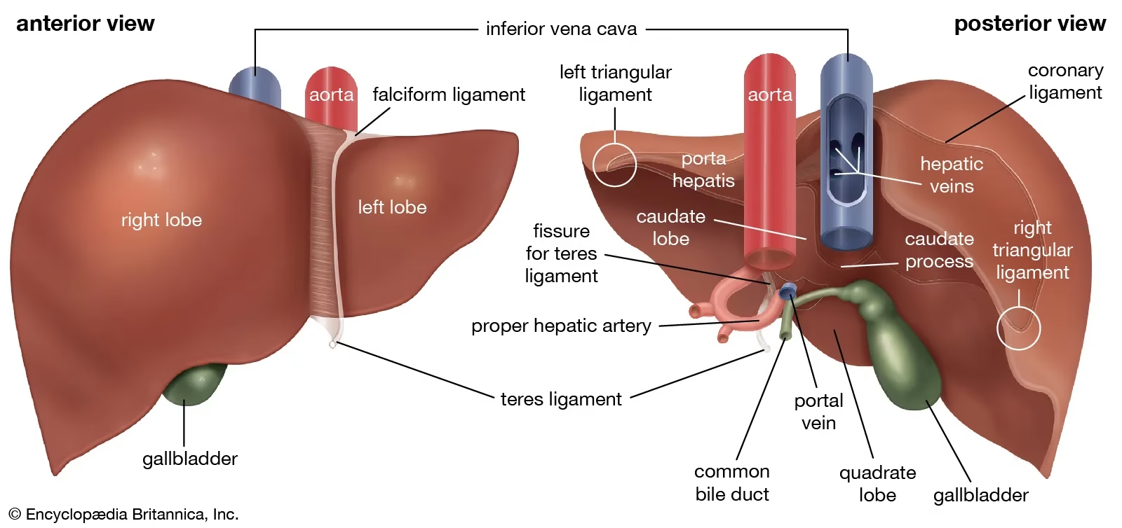
(A) Lobes of the Liver
The liver is divided into four lobes based on external anatomy:
-
Right Lobe:
- The largest lobe.
- It performs the majority of the liver’s metabolic and detoxification functions.
- Contains the gallbladder fossa (a depression where the gallbladder is located).
-
Left Lobe:
- Smaller than the right lobe.
- It extends towards the left side of the body.
-
Caudate Lobe:
- A small lobe is located on the posterior surface of the liver.
- Lies between the inferior vena cava and the ligamentum venosum.
- Functionally related to both the right and left lobes.
-
Quadrate Lobe:
- Located near the gallbladder and bile duct.
- It plays a role in bile secretion.
(B) Surfaces of the Liver
-
Diaphragmatic Surface:
- The smooth, convex surface that faces the diaphragm.
- In direct contact with the diaphragm, separating it from the heart and lungs.
- Covered by the peritoneum, except in the bare area (where it is attached directly to the diaphragm).
-
Visceral Surface:
- The inferior surface faces the abdominal organs.
- Contains impressions for the stomach, right kidney, colon, and duodenum.
- The porta hepatis (entry point for blood vessels, nerves, and bile ducts).
(C) Ligaments of the Liver
The liver is held in position by several ligaments, which are foldings of the peritoneum:
-
Falciform Ligament:
- Divides the liver into the right and left lobes.
- Attaches the liver to the anterior abdominal wall.
-
Round Ligament (Ligamentum Teres Hepatis):
- A remnant of the fetal umbilical vein.
- Runs along the inferior edge of the falciform ligament.
-
Coronary Ligament:
- Attaches the superior part of the liver to the diaphragm.
-
Triangular Ligaments (Right and Left):
- Formed by extensions of the coronary ligament.
- Help secure the liver in place.
Internal Structure of the Liver
The internal structure of the liver consists of specialized cells, blood vessels, and functional units called hepatic lobules.
(A) Hepatic Lobules: The Functional Unit of the Liver
- The liver comprises millions of hexagonal-shaped hepatic lobules (~1-2 mm in diameter).
- Each lobule is centred around a central vein and surrounded by a portal triad.
- The lobules comprise hepatocytes (liver cells), which carry out the liver’s metabolic, detoxification, and bile-producing functions.
(B) Portal Triad (Glisson’s Triad)
Each corner of the lobule contains a portal triad, which consists of three major structures:
- Hepatic Artery Branch:
- Brings oxygen-rich blood to the liver from the heart.
- Hepatic Portal Vein Branch:
- Carries nutrient-rich blood from the intestines for processing.
- Bile Duct:
- Collects bile produced by hepatocytes and sends it to the gallbladder and duodenum.
(C) Sinusoids and Blood Flow
- Hepatic sinusoids are specialized capillaries lined with endothelial and Kupffer cells.
- Blood from the hepatic artery and portal vein enters the sinusoids, exchanging nutrients and oxygen with hepatocytes.
- After processing, blood drains into the central vein, the hepatic veins, and finally into the inferior vena cava, returning to the heart.
(D) Kupffer Cells (Liver Macrophages)
- Kupffer cells are immune cells located in the sinusoids.
- Functions:
- Destroy bacteria, toxins, and dead red blood cells.
- Help in liver detoxification and immune response.
Blood Supply of the Liver
The liver has a unique dual blood supply, receiving blood from two major sources:
-
Hepatic Artery (25%)
- Carries oxygenated blood from the aorta (via the celiac trunk).
-
Hepatic Portal Vein (75%)
- Carries nutrient-rich blood from the stomach, intestines, pancreas, and spleen.
- This allows the liver to process absorbed nutrients and detoxify harmful substances before blood enters general circulation.
Venous Drainage:
- Blood from the liver drains into the central vein of each lobule.
- Central veins merge to form the hepatic veins, which empty into the inferior vena cava, returning blood to the heart.
Biliary System: Bile Production and Flow
The biliary system is responsible for producing, storing, and transporting bile.
- Bile Canaliculi: Tiny ducts within the lobules where hepatocytes secrete bile.
- Bile Ductules → Hepatic Ducts: Small bile canaliculi drain into bile ducts.
- Common Hepatic Duct: Formed by the right and left hepatic ducts.
- Gallbladder Storage: The common hepatic duct joins the cystic duct from the gallbladder to form the common bile duct, which empties into the duodenum.
Liver Function Based on Its Structure
| Liver Structure | Function |
|---|---|
| Hepatic Lobules | Main functional unit for metabolism, detoxification, and bile production. |
| Hepatocytes | Perform digestion, detoxification, and protein synthesis. |
| Sinusoids | Allow nutrient and oxygen exchange between blood and hepatocytes. |
| Kupffer Cells | Remove pathogens, toxins, and old RBCs from the blood. |
| Portal Triad | Supplies blood and collects bile. |
| Biliary System | Produces, stores, and releases bile for digestion. |