Principle
- The principle of AFB staining hinges on the unique properties of mycobacterial cell walls.
- These bacteria contain a high concentration of mycolic acids, which make their cell walls impervious to most dyes.
- The AFB staining method relies on a potent red dye called carbolfuchsin, which penetrates the cell wall.
- Once inside, the dye is trapped by the thick lipid layer, making it resistant to decolorization by acid-alcohol.
- This resistance is what makes acid-fast bacteria “stand out” under the microscope, even after being subjected to harsh decolorizing agents.
Requirements
-
Microscope: A compound light microscope with an oil immersion lens (100x) to clearly view the acid-fast bacilli.
-
Glass slides: Clean slides for preparing bacterial smears.
-
Heat source: For fixing the smear and aiding in dye penetration.
-
Inoculating loop: To transfer the bacterial sample onto the slide.
-
Staining rack: To hold the slides during the staining process.
Reagents
-
Carbolfuchsin (Primary stain): This powerful red dye is the star of the show, able to bind to the bacteria’s waxy cell walls.
-
Acid-alcohol (Decolorizer): A mixture of alcohol and hydrochloric acid that removes the dye from non-acid-fast bacteria, leaving acid-fast bacilli untouched.
-
Methylene blue (Counterstain): Provides a contrasting blue color to non-acid-fast cells, making the acid-fast bacteria even easier to spot.
Specimen
-
Clinical Specimens: Sputum, urine, blood, cerebrospinal fluid, and tissue biopsies are the primary samples used in AFB testing. These are often collected from patients suspected of having tuberculosis or other mycobacterial infections.
-
Smear Preparation: A small amount of the sample is transferred onto a glass slide and air-dried before being heat-fixed to ensure the bacteria stay in place during the staining process.
Procedure
-
Prepare the smear: Take a small drop of the clinical specimen and spread it evenly on the slide. Allow the smear to air dry before heat-fixing it by passing the slide through a flame.
-
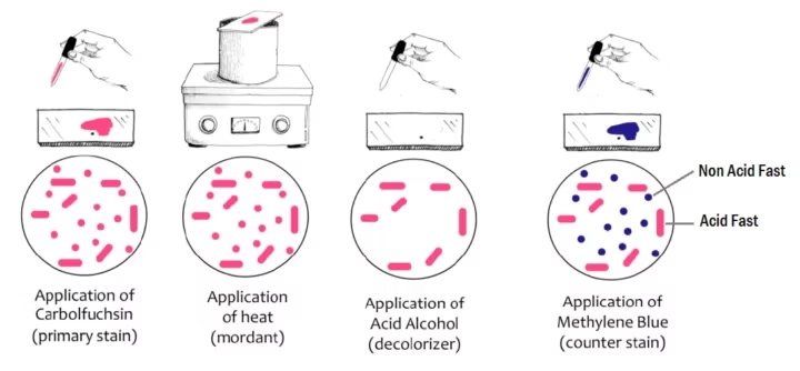
Stain with carbolfuchsin: Place a few drops of carbolfuchsin onto the smear. Gently heat the slide for about 5 minutes. This step ensures the dye penetrates the bacteria’s tough, waxy walls.
-
Decolorization: After cooling the slide, rinse it with water to remove excess dye. Then, apply acid-alcohol for 1-2 minutes. This will strip the red stain from all non-acid-fast bacteria, leaving the acid-fast bacteria red.
-
Counterstain with methylene blue: Rinse the slide with water and apply methylene blue for 1-2 minutes. The blue dye will color the non-acid-fast cells and the background, providing contrast.
-
Rinse, dry, and examine: After rinsing off excess counterstain, gently blot the slide dry. Allow it to air-dry completely before examining it under a microscope.
-
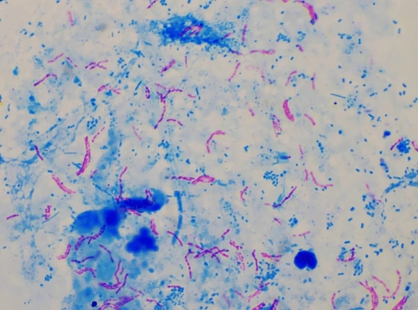
Examine under the microscope: Using an oil immersion lens, look for red, rod-shaped bacteria—these are your acid-fast bacilli! The rest of the cells will appear blue, allowing for easy differentiation.
Results
-
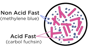
Acid-Fast Bacilli (AFB): These bacteria will appear bright red or pink against the blue background, indicating that they’ve retained the carbolfuchsin stain despite the acid-alcohol treatment.
-
Non-Acid-Fast Cells: These will appear blue, as they lose the carbolfuchsin stain and retain the methylene blue counterstain.
Applications
-
Diagnosing Tuberculosis (TB): AFB staining is one of the most effective ways to detect Mycobacterium tuberculosis in sputum, helping to confirm a TB diagnosis quickly and accurately.
-
Identifying Leprosy: AFB staining is used to detect Mycobacterium leprae in skin biopsies and nasal secretions, offering a crucial diagnostic tool for leprosy.
-
Mycobacterial Infections Beyond TB: The technique can also identify other mycobacteria, such as Mycobacterium avium and Mycobacterium intracellulare, which cause infections in immunocompromised individuals.
-
Environmental Testing: AFB staining is used to detect mycobacteria in water and soil, which can help prevent environmental contamination and subsequent infections.
-
Research and Surveillance: In scientific research, AFB staining aids in studying the structure and behavior of Mycobacterium species, supporting advancements in treatment and vaccine development.