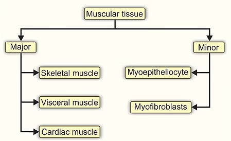
Types of muscular tissue
Types
There are the following three types of muscles:
1. Skeletal or striated muscle
2. Cardiac muscle
3. Smooth or visceral muscle
Apart from the above three types, there are contractile cells functioning as single cell units, namely,
1. Myoepithelial cells (found in association with secretory acini)
2. Myofibroblasts (involved in wound healing)
3. Myoid cells (found around seminiferous tubules)
Skeletal Muscle
General feature
The skeletal muscle fibres are elongated, cylindrical, multinucleated cells whose length varies from a few millimetres to 35 cm
and width from 10 μm to 100 μm.
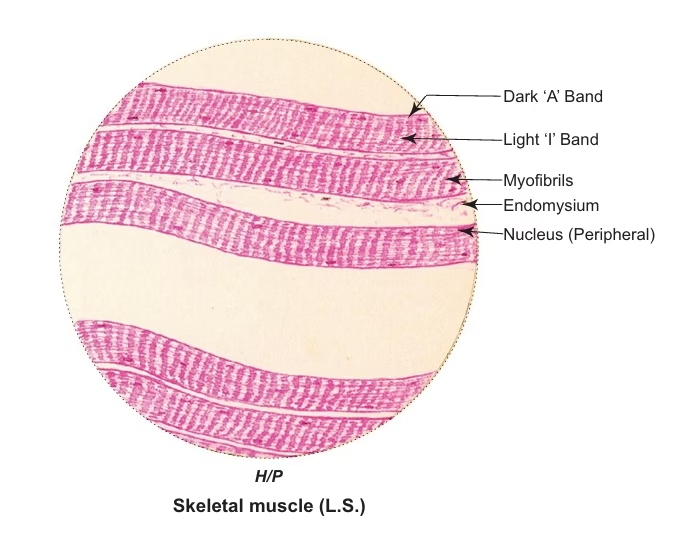
Structure of skeletal muscle fibre
Light Microscopic (LM) Observation
- Each muscle fibre is an elongated, unbranched, cylindrical cell.
- It has many flat nuclei located just beneath the sarcolemma.
- It shows cross striations of alternate dark (A) and light (I) bands with Z line intersecting I band.
- Each muscle fibre is made of compactly packed, long, cylindrical myofibrils in the sarcoplasm arranged parallel to the long
axis.
Types of skeletal muscle fibres
There are three main types of skeletal muscle fibres, each adapted for different functions:
-
Type I (Slow-Twitch Fibres):
-
Function: Primarily used for endurance and activities that require long-duration efforts (e.g., long-distance running).
-
Characteristics: These fibres contract slowly and are highly resistant to fatigue. They have a high amount of mitochondria and myoglobin, making them efficient at using oxygen for energy production.
-
Appearance: Red in colour due to the high myoglobin content.
-
-
Type IIa (Fast-Twitch, Oxidative Fibers):
-
Function: Used for moderate-intensity activities that require both endurance and strength (e.g., swimming or middle-distance running).
-
Characteristics: These fibers contract quickly and are somewhat resistant to fatigue. They rely on both aerobic (oxygen) and anaerobic (without oxygen) metabolism for energy.
-
Appearance: Pink in color due to a mix of myoglobin and glycogen.
-
-
Type IIb (Fast-Twitch, Glycolytic Fibers):
-
Function: Used for short bursts of high-intensity activities (e.g., sprinting, weightlifting).
-
Characteristics: These fibers contract rapidly and with great force but tire quickly. They rely on anaerobic metabolism and use glycogen for energy.
-
Appearance: White in color due to low myoglobin content.
-
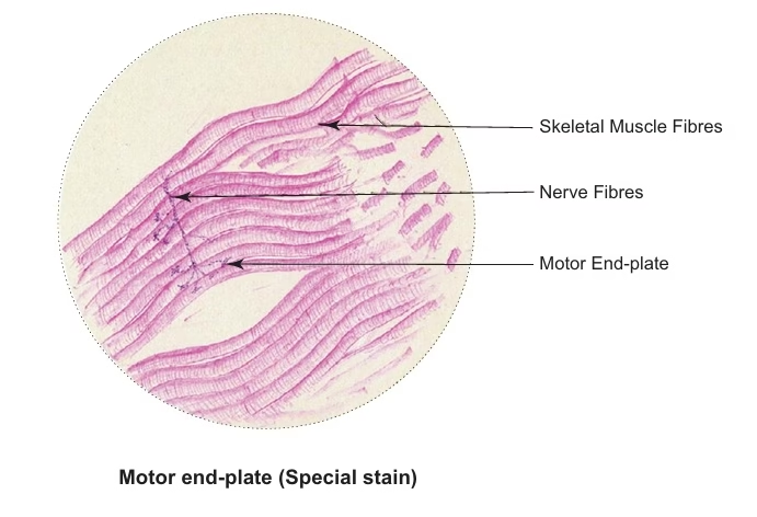
Motor end plates
- Skeletal muscle is richly innervated by myelinated motor nerves (axons).
- At the site of innervation, each axon divides into many terminal twigs that end in dilated bulbs called bouton terminals on the muscle surface.
- It is this specialised site where the axon terminates on the surface of skeletal muscle, that is called motor end-plate or neuromuscular junction.
- It is the site where the impulses from the axon are transmitted to the skeletal muscle fibres.
- The neurotransmitter released at the site is acetylcholine.
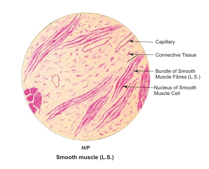
Smooth muscle
- Smooth muscle fibres are elongated, spindle-shaped cells measuring 30 μm in length in blood vessels to 200– 500 μm in the pregnant
uterus. - They are nonstriated (smooth), involuntary and supplied by the autonomic nervous system.
- Adjacent smooth muscle cells are in contact with each other through gap junctions, which help to transmit the electric impulses from one cell to another, resulting in simultaneous contraction of the entire muscle.
- Smooth muscle fibres are found in the walls of hollow viscera (viz., G.I.T., blood vessels, ureter, uterine tube, vas deferens, etc.) and also occur as separate entities like arrector pili, ciliaris, sphincter pupillae, etc.
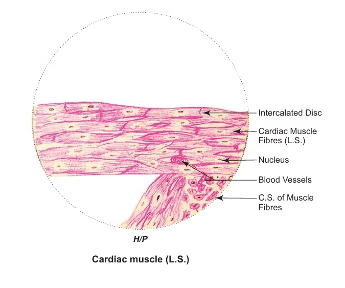
Cardiac muscle
- Cardiac muscle shows many structural and functional characteristics intermediate between those of skeletal and smooth muscle.
- Though it exhibits cross striations, like skeletal muscle, it is involuntary and contracts automatically like smooth muscle.
- Cardiac muscle fibres are shorter than skeletal muscle fibres and show a branching pattern.
- They have one or two nuclei placed in the centre.
- The most striking feature of cardiac muscle is the presence of darkly staining transverse lines across the fibres called intercalated discs which are specialised cell junctions between the ends of adjacent muscle fi bres.
- These cell junctions (gap junctions and desmosomes) provide a mechanism by which the contractile stimuli pass from one cell to another, causing the adjacent cell to contract simultaneously.
- Thus, cardiac muscle acts as a functional syncytium.
- The conducting system of the heart (SA node, AV node, bundle of His and Purkinje fibres) is made of modified cardiac
muscle fibres, which are thicker, larger and contain few myofibrils and are found just deep to the endocardium.
Applied Histology
-
Skeletal Muscle Disorders:
-
Muscular Dystrophy: Progressive muscle weakness; histology shows muscle fiber degeneration and replacement with fat and connective tissue.
-
Rhabdomyolysis: Muscle breakdown, releasing myoglobin; histology shows muscle fiber necrosis.
-
Strain and Repair: Muscle injury causes fiber rupture; satellite cells repair muscle, but extensive injury leads to scar tissue.
-
-
Cardiac Muscle Disorders:
-
Cardiomyopathies: Heart muscle diseases, such as dilated or hypertrophic cardiomyopathy, show fiber enlargement and disarray.
-
Myocardial Infarction: Heart attack causes muscle death, replaced by scar tissue.
-
-
Smooth Muscle Disorders:
-
Hypertension: Arterial smooth muscle thickens, narrowing blood vessels.
-
Asthma: Smooth muscle thickens in airways, causing bronchoconstriction.
-
Irritable Bowel Syndrome (IBS): Abnormal smooth muscle contraction in the intestines.
-
-
Regeneration and Repair:
-
Satellite Cells: Muscle stem cells that repair damage by fusing with fibers, though regeneration declines with age.
-
Fibrosis: Chronic injury or aging leads to muscle fibrosis, reducing function.
-
-
Exercise Adaptation:
-
Hypertrophy: Muscle fiber size increases with strength training.
-
Atrophy: Muscle fibers shrink due to disuse or disease.
-
Fiber Type Transformation: Endurance training can convert fast-twitch fibers to more fatigue-resistant types.
-
-
Muscle Disease Diagnosis:
-
Histology helps diagnose muscle diseases like dystrophies, myositis, autoimmune conditions, and neurogenic disorders by revealing inflammation, fiber degeneration, or changes in muscle structure.
-