
Introduction
- Cell biology is often called the basic unit of life. Every living organism, from the simplest bacteria to the most complex human being, is built from cells.
- In the human body, trillions of cells work together in a coordinated way to carry out all essential processes of life, such as energy production, growth, repair, communication, defence, and reproduction.
- Cells are not just small “building blocks,” but highly organized systems with specialized parts that function like miniature factories.
- Human cells are eukaryotic, which means they have a true nucleus enclosed by a membrane and contain many membrane-bound organelles.
- These organelles divide the cell into different compartments, each carrying out a specific job.
- This compartmentalisation is what makes eukaryotic cells efficient and complex compared to prokaryotic cells (like bacteria).
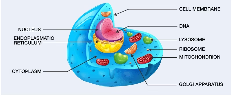
Even though there are over 200 different types of human cells (such as nerve cells, muscle cells, blood cells, and epithelial cells), all of them share the same basic structure:
- The cell membrane (plasma membrane) – the protective barrier.
- The cytoplasm – the fluid matrix that holds organelles.
- The nucleus – the control center containing genetic material.
- Organelles – the “machines” that perform different tasks within the cell.
In this essay, we will discuss the structure of a human cell in detail, with special attention to the components of the cell, intracellular organelles, and the cell membrane.
General Structure of a Human Cell
Cell Membrane (Plasma Membrane)
- The outermost covering of the cell is the cell membrane, also known as the plasma membrane.
- It is a thin, flexible barrier that separates the cell’s internal contents from its external environment.
- The cell membrane is about 7–10 nanometers thick and is selectively permeable, which means it controls what substances can enter or leave the cell.
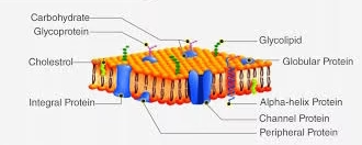
The structure of the cell membrane is best explained by the Fluid Mosaic Model (Singer and Nicolson, 1972). According to this model:
- The membrane is made of a phospholipid bilayer – two layers of phospholipids with hydrophilic (water-attracting) heads facing outward and hydrophobic (water-repelling) tails facing inward.
- Proteins are embedded in this bilayer, some spanning across the membrane (integral proteins) and others attached to the surface (peripheral proteins).
- Cholesterol molecules are scattered within the bilayer, making the membrane flexible and stable.
- Carbohydrate chains are attached to proteins (glycoproteins) or lipids (glycolipids), and they help in cell recognition and communication.
Functions of the Cell Membrane:
- Protection: Acts as a barrier between the cell and its environment.
- Selective Permeability: Controls the entry of nutrients, gases, and the exit of waste products.
- Transport: Facilitates passive transport (diffusion, osmosis) and active transport (ATP-driven pumps, endocytosis, exocytosis).
- Communication: Membrane proteins act as receptors for hormones, neurotransmitters, and other signaling molecules.
- Recognition: Glycoproteins and glycolipids help the immune system distinguish between “self” and “non-self” cells.
- Support: Provides structural support and helps cells attach to one another to form tissues.
Cytoplasm
The cytoplasm is the jelly-like material inside the cell, filling the space between the nucleus and the plasma membrane. It consists of:
- Cytosol: The fluid part, made up of water, salts, and organic molecules.
- Organelles: Specialized structures with specific functions.
- Cytoskeleton: A network of fibers that gives shape and allows movement.
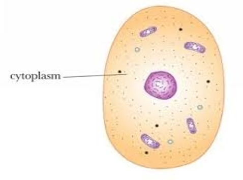
The cytoplasm is the site of many metabolic reactions, such as glycolysis, and also serves as the medium in which organelles are suspended.
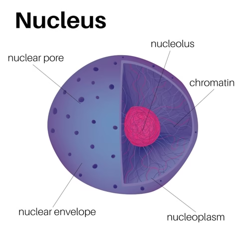
Nucleus
The nucleus is the largest and most important organelle in human cells, often called the control center. It is surrounded by the nuclear envelope, a double membrane with pores that regulate the movement of molecules in and out.
Inside the nucleus:
- Chromatin: DNA wrapped around histone proteins, containing genetic instructions. During cell division, chromatin condenses into chromosomes.
- Nucleolus: A dense body that produces ribosomal RNA (rRNA) and assembles ribosome subunits.
- Nucleoplasm: The fluid inside the nucleus.
Functions of the Nucleus:
- Stores and protects DNA.
- Directs protein synthesis by producing messenger RNA (mRNA).
- Controls growth, metabolism, and reproduction.
- Coordinates cell division (mitosis and meiosis).
Intracellular Organelles and Their Functions
Now, let us look at the major organelles in detail:
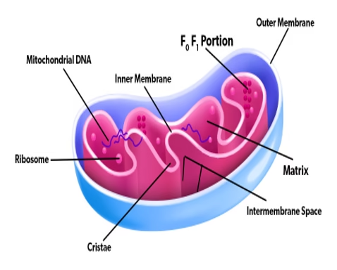
Mitochondria – The Powerhouse
- Double-membrane organelle; inner membrane has folds called cristae.
- Contains its own DNA and ribosomes, meaning it can produce some of its own proteins.
- Site of aerobic respiration: converts glucose and oxygen into ATP (adenosine triphosphate).
Functions:
- Energy production (ATP).
- Regulation of metabolism.
- Involvement in cell death (apoptosis).
- Calcium storage.
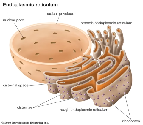
Endoplasmic Reticulum (ER) – The Transport System
A network of membranes connected to the nuclear envelope.
- Rough ER (RER): Covered in ribosomes. Synthesizes proteins that are secreted from the cell or embedded in membranes.
- Smooth ER (SER): Lacks ribosomes. Synthesizes lipids, cholesterol, steroids, detoxifies drugs and poisons, and stores calcium.
Functions:
- RER: Protein synthesis and transport.
- SER: Lipid synthesis, detoxification, and regulation of calcium.
Ribosomes – The Protein Factories
- Composed of rRNA and proteins.
- Found free in the cytoplasm or attached to the RER.
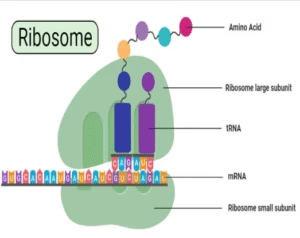
Functions:
- Translate mRNA into proteins (protein synthesis).
- Free ribosomes make proteins used within the cell.
- Attached ribosomes make proteins exported out of the cell.
Golgi Apparatus – The Post Office
- Stack of flattened sacs (cisternae).
- Receives proteins and lipids from ER.
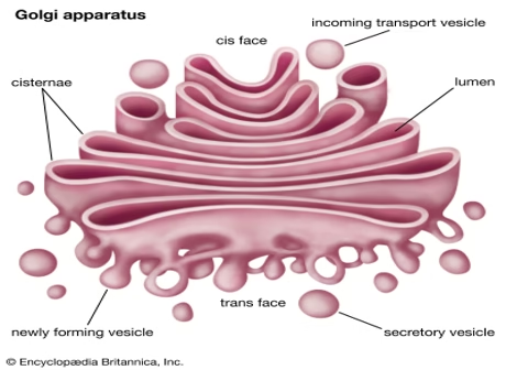
Functions:
- Modifies proteins and lipids (adds sugars to make glycoproteins).
- Sorts and packages them into vesicles.
- Produces lysosomes.
- Secretes substances like hormones and enzymes.
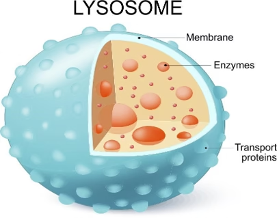
Lysosomes – The Waste Disposal System
- Membrane-bound sacs containing digestive enzymes.
- Spherical, membrane-bound organelles containing hydrolytic (digestive) enzymes.
- They are surrounded by a single membrane that prevents enzyme leakage.
- Formed from the Golgi apparatus and found mainly in animal cells.
Functions:
-
Intracellular Digestion: Break down worn-out organelles, food particles, and engulfed pathogens.
-
Autophagy: Digests and recycles the cell’s own damaged organelles.
-
Autolysis: Causes the self-destruction of damaged or dead cells (“suicidal bags”).
-
Defence: Destroy invading bacteria and viruses through enzymatic action.
-
Waste Removal: Help in clearing cellular debris and maintaining cell cleanliness.
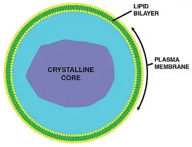
Peroxisomes – The Detoxifiers
- Contain oxidative enzymes.
- Small, spherical, membrane-bound organelles containing oxidative enzymes such as catalase and oxidase.
- They have a single membrane and a granular or crystalline core.
- Abundant in liver and kidney cells, where detoxification and fatty acid breakdown occur.
Functions:
- Break down fatty acids.
- Detoxify alcohol and harmful substances.
- Neutralise hydrogen peroxide (H₂O₂) into water and oxygen.
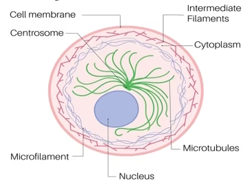
Cytoskeleton – The Framework
- Protein filaments that provide structure.
- Microtubules: Hollow tubes that transport organelles and form spindle fibers.
- Microfilaments: Thin filaments for movement and support.
- Intermediate filaments: Provide mechanical strength.
Functions:
- Maintains cell shape.
- Assists in cell movement.
- Helps in transport of organelles and vesicles.
- Essential in cell division.
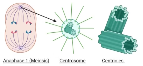
Centrosome and Centrioles
- Found near the nucleus.
- The centrosome contains two centrioles arranged at right angles.
- The centrosome is located near the nucleus and contains two centrioles arranged at right angles.
- Each centriole is made up of nine sets of microtubule triplets arranged in a cylindrical pattern.
- They lack a surrounding membrane and play a key role in spindle formation during cell division.
Functions:
-
Spindle Formation: Help in forming the spindle fibers during cell division.
-
Chromosome Movement: Guide the movement of chromosomes to opposite poles.
-
Microtubule Organization: Act as the main center for organizing microtubules in the cell.
-
Cilia and Flagella Formation: Centrioles give rise to basal bodies that form cilia and flagella.
Vacuoles
- Membrane-bound sacs for storage.
- Small in animal cells; large central vacuole in plant cells.
- Membrane-bound sacs filled with fluid or stored substances.
- Surrounded by a single membrane called the tonoplast.
- Contain water, nutrients, salts, and waste materials.
- Large and central in plant cells; small and temporary in animal cells.
Functions:
-
Storage: Store water, nutrients, salts, and waste products.
-
Osmoregulation: Maintain water balance and internal pressure (turgor pressure) in plant cells.
-
Waste Disposal: Isolate harmful materials and metabolic waste.
-
Support: Provide rigidity to plant cells by maintaining turgidity.
-
Digestion: Contain enzymes that help in breaking down macromolecules in some cells.
Other Structures
- Cilia and Flagella: Extensions of the plasma membrane that aid in movement (e.g., cilia in respiratory tract move mucus).
- Vesicles: Small sacs used to transport substances.
Cell Membrane
- The cell membrane, also called the plasma membrane, is the thin, flexible outer covering of the cell.
- It separates the inside of the cell (intracellular environment) from the outside (extracellular environment).
- It is not just a passive boundary — it actively controls what enters and exits the cell, helps in communication, and provides structural support.
- The cell membrane is about 7–10 nanometers thick and is present in all living cells.
- In human biology, it plays a vital role because cells constantly interact with their environment, absorb nutrients, release wastes, and respond to chemical signals.
Structure of the Cell Membrane
The structure of the cell membrane is best explained by the Fluid Mosaic Model (Singer and Nicolson, 1972).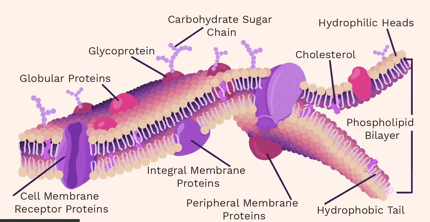
- Phospholipid Bilayer
- The cell membrane is mainly made up of phospholipids, which are special fat molecules.
- Each phospholipid has:
- A hydrophilic head (water-loving) made of phosphate.
- Two hydrophobic tails (water-fearing) made of fatty acids.
- In the membrane, phospholipids arrange themselves in two layers (bilayer):
- Heads face outward toward water (both inside and outside the cell).
- Tails face inward, away from water.
- This arrangement makes the membrane semi-permeable (some substances can pass, others cannot).
- Proteins
Proteins are embedded in the phospholipid bilayer, forming a “mosaic” pattern.
- Integral proteins: Span across the membrane. They act as channels, pumps, or transporters.
- Peripheral proteins: Found on the inner or outer surface. They act as enzymes or anchors for the cytoskeleton.
- Cholesterol
- Scattered within the bilayer.
- Provides stability by preventing the membrane from becoming too stiff or too fluid.
- Carbohydrates
- Attached to proteins (glycoproteins) or lipids (glycolipids).
- Extend outward from the cell surface.
- Important for cell recognition and communication. (e.g., immune cells identifying foreign invaders).
Functions of the Cell Membrane
The plasma membrane is dynamic and multifunctional:
- Protection
- Acts as a barrier separating the cell’s internal environment from the external environment.
- Protects organelles and vital molecules inside the cell.
- Selective Permeability
- Controls the movement of substances in and out of the cell.
- Allows essential nutrients and oxygen to enter.
- Removes waste products like carbon dioxide.
- Prevents harmful substances from entering.
- Transport
There are several ways substances cross the membrane:
- Passive Transport (no energy):
- Diffusion: Movement of molecules from high to low concentration (e.g., oxygen).
- Osmosis: Movement of water across the membrane.
- Facilitated diffusion: Molecules pass through protein channels (e.g., glucose).
- Active Transport (requires energy/ATP):
- Protein pumps move substances against their concentration gradient.
- Endocytosis: Cell engulfs large particles into vesicles.
- Exocytosis: Cell releases large molecules by vesicles fusing with the membrane.
- Communication
- Membrane proteins act as receptors.
- Receptors detect signals such as hormones, neurotransmitters, and chemical messengers.
- This allows cells to respond to changes in the environment.
- Cell Recognition
- Glycoproteins and glycolipids act as “identity markers.”
- Helps the immune system distinguish between the body’s own cells and foreign invaders (pathogens).
- Structural Support
- Anchors the cytoskeleton inside the cell, giving it shape.
- Connects with other cells via cell junctions to form tissues.
Specialized Cell Membrane Features
In some human cells, the plasma membrane has specialized structures:
- Microvilli: Tiny finger-like projections that increase surface area for absorption (e.g., intestinal cells).
- Cilia: Hair-like structures that move substances along the surface (e.g., respiratory tract cells).
- Flagella: Tail-like structure that helps movement (e.g., sperm cells).