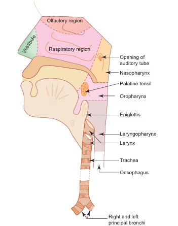
Structure
Histologically, based on the lining epithelium, the nasal cavity is divided into three regions.
1. Vestibule
-
It is the anterior dilated part of the nasal cavity and is lined by skin.
-
It contains thick short hairs called vibrissae, which filter large particles from the inspired air.
2. Olfactory region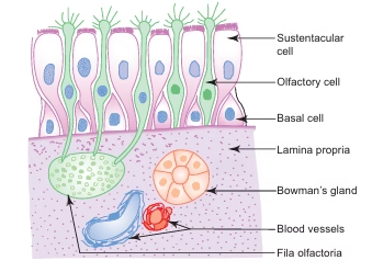
-
This region is confined to a smaller area and is present in the roof and adjoining parts of the nasal septum and lateral wall of the nasal cavity.
-
It is covered by a thick yellow olfactory mucosa which functions as the organ of smell.
-
The olfactory mucosa is composed of a thick olfactory epithelium and the underlying connective tissue called lamina propria.
-
The lamina propria contains serous glands (bowman’s glands), bundles of olfactory nerves (fila olfactoria), blood vessels, and lymphatics.
-
The secretion of bowman’s glands moistens the olfactory epithelium and acts as a solvent for odoriferous substances, thereby stimulating the olfactory cells.
-
The olfactory epithelium consists of the following three types of cells.
(a) Olfactory cells
-
These are modified bipolar nerve cells.
-
Each cell has a cell body, a dendrite, and an axon.
-
The cell bodies contain round nuclei and are found at different levels, forming a broad zone of round nuclei.
-
The dendrites extend to the surface and end in knob-like olfactory vesicles from which nonmotile olfactory hairs arise.
-
The axons pass into the lamina propria and form bundles of olfactory nerves (fila olfactoria).
(b) Sustentacular cells
-
These are tall columnar supporting cells.
-
They bear microvilli on their free surface.
-
Each cell has a broad superficial part with an oval nucleus and a slender deep part attached to the basement membrane.
-
Their nuclei lie near the free surface at the same level, forming a narrow zone of oval nuclei.
-
The presence of yellow lipofuscin pigments suggests a phagocytic function.
(c) Basal cells
-
These cells are conical in shape.
-
They do not reach the surface.
-
They give rise to other cell types.
3. Respiratory region
-
It occupies the remaining area of the nasal cavity.
-
It is covered by respiratory mucosa, which is pink in color and thinner than the olfactory mucosa.
-
The respiratory mucosa is firmly adherent to the underlying periosteum or perichondrium.
-
It consists of respiratory epithelium and underlying lamina propria.
-
The respiratory epithelium is pseudostratified ciliated columnar epithelium with goblet cells.
-
The lamina propria contains mixed glands, lymphatic nodules, and rich vascular plexuses.
-
The respiratory epithelium includes the following cell types.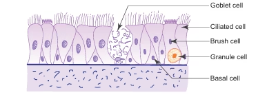
(a) Ciliated cells
-
These are columnar cells with cilia on their free surface.
-
They are the most abundant cell type.
-
The cilia beat towards the pharynx.
(b) Goblet cells
(c) Brush cells
(d) Basal cells
-
These are small pyramidal cells.
-
They do not reach the surface.
-
They give rise to other cell types.
(e) Granule cells
Pharynx
General features
-
Pharynx is a fibromuscular tube extending from the base of the skull to the level of the sixth cervical vertebra, where it becomes continuous with the oesophagus.
-
It lies behind the nasal cavity forming the nasopharynx, behind the oral cavity forming the oropharynx, and behind the larynx forming the laryngopharynx.
Structure
1. Mucosa
-
It consists of epithelium and lamina propria.
-
The epithelium is pseudostratified ciliated columnar type in the nasopharynx.
-
The epithelium is stratified squamous type in the oropharynx and laryngopharynx.
-
Aggregation of lymphatic nodules in the lamina propria of the posterior wall and around the opening of the auditory tube in the nasopharynx forms the pharyngeal tonsil and tubal tonsils respectively.
-
The palatine tonsil in the lateral wall of the oropharynx and the lingual tonsil in the pharyngeal part of the tongue are described under the lymphatic system.
2. Submucosa
3. Muscle coat
-
This layer is composed of skeletal muscle.
-
The muscle fibers are arranged into an inner longitudinal layer and an outer circular layer.
-
The circular layer is formed by the constrictor muscles of the pharynx.
4. Adventitia
- It is formed by fibroelastic connective tissue (buccopharyngeal fascia).
Larynx
General features
-
Larynx is a specialised organ responsible for the production of voice.
-
It houses the vocal cords.
-
Above, it opens into the laryngopharynx, and below it is continuous with the trachea.
-
It has a cartilaginous framework made up of nine cartilages, consisting of three paired and three unpaired cartilages.
-
These cartilages are connected to each other by membranes and ligaments and are moved by skeletal muscles.
-
The cartilages are either hyaline or elastic in nature.
-
The hyaline cartilages include:
-
Thyroid cartilage (unpaired)
-
Cricoid cartilage (unpaired)
-
Arytenoid cartilages (paired)
-
The elastic cartilages include:
Structure
-
Most of the larynx is lined by pseudostratified ciliated columnar epithelium with goblet cells.
-
In areas subjected to friction or contact with food, it is lined by stratified squamous epithelium.
-
Examples of such areas include the anterior surface and upper part of the posterior surface of the epiglottis and the vocal folds.
-
The lamina propria may contain seromucous glands and lymphatic tissue.
Trachea
General features
-
Trachea is a flexible fibroelastic cartilaginous tube that allows expansion in width and extension in length during inspiration.
-
It measures about 10 cm in length and 2 cm in width.
-
It extends from the lower border of the cricoid cartilage at the level of c6 to its bifurcation at the level of t4 into right and left bronchi.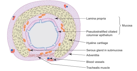
-
It is also called the windpipe and serves as the airway through which respiratory air is transported.
-
It is supported by 16–20 c-shaped rings of hyaline cartilage that keep the lumen patent.
-
The free posterior ends of the c-shaped cartilages are connected by a band of smooth muscle called trachealis and a fibroelastic ligament.
-
The ligament prevents overdistension of the lumen, while contraction of the muscle reduces the diameter during coughing, increasing the velocity of expired air for cleaning the air passages.
Structure
The trachea is composed of four coats, similar to the general plan of the respiratory tract.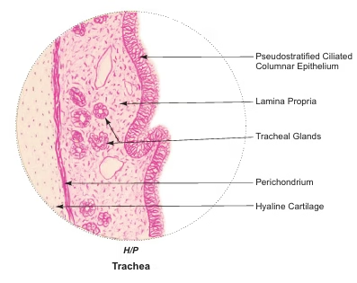
1. Mucosa
-
It is composed of epithelium and lamina propria.
-
The epithelium is thick and consists of pseudostratified ciliated columnar epithelium with goblet cells.
-
The lamina propria is made of fibroelastic vascular connective tissue with longitudinally oriented elastic fibers.
-
Lymphocytes and mast cells are abundant in the lamina propria.
2. Submucosa
-
It is formed by loose connective tissue located deep to the lamina propria.
-
It contains mixed glands.
-
There is no clear demarcation between the lamina propria and the submucosa.
3. Cartilage and smooth muscle layer
4. Adventitia
Lungs
General features
-
Lungs are the principal organs of respiration and are situated in the thoracic cavity on either side of the mediastinum.
-
Each lung is conical in shape and is covered by visceral pleura.
-
Lungs contain the terminal parts of the bronchial tree, including intrapulmonary bronchi, bronchioles, respiratory bronchioles, and lung parenchyma consisting of alveolar ducts and alveoli, along with blood vessels.
Structure
-
The lining epithelium of the bronchial tree gradually decreases in thickness towards the distal parts.
-
Glands and goblet cells also decrease gradually and disappear distally.
-
Cartilaginous support decreases, while elastic fibers increase towards the periphery.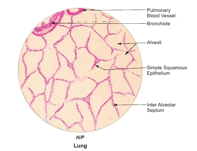
1. Intrapulmonary bronchus (secondary and tertiary bronchi)
-
Each principal bronchus divides into secondary (lobar) bronchi, which further divide into tertiary (segmental) bronchi.
-
The following layers are seen from inside to outside.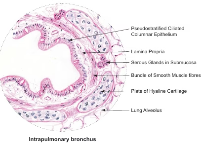
(a) Mucosa
-
It consists of epithelium and lamina propria.
-
The epithelium is pseudostratified ciliated columnar with few goblet cells.
-
The lamina propria is rich in longitudinally arranged elastic fibers.
-
The mucosa is thrown into folds due to contraction of smooth muscle.
(b) Smooth muscle layer
(c) Submucosa
(d) Cartilage layer and adventitia
2. Bronchiole
-
Bronchioles are formed by repeated division of tertiary bronchi.
-
Each bronchiole enters a pulmonary lobule and divides into 5–7 terminal bronchioles.
-
Terminal bronchioles have a diameter of less than 1 mm.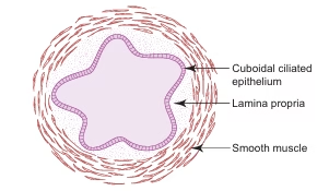
-
Bronchioles show the following features:
-
Simple columnar or cuboidal ciliated epithelium.
-
Goblet cells are absent and replaced by clara cells.
-
Clara cells secrete a protective glycoprotein.
-
Thick smooth muscle layer under autonomic control.
-
Many elastic fibers.
-
Absence of glands and cartilage.
3. Respiratory bronchiole
-
Respiratory bronchioles arise from terminal bronchioles.
-
They form the transitional zone between conducting and respiratory portions.
-
Their walls are interrupted by alveolar outpouchings.
-
The lining epithelium between alveoli is ciliated cuboidal with clara cells.
-
They divide distally into alveolar ducts.
4. Alveolar duct
-
Alveolar ducts arise from respiratory bronchioles.
-
They are lined by squamous epithelial cells supported by smooth muscle and fibroelastic tissue.
-
Numerous alveoli open into the ducts, giving a knob-like appearance in sections.
-
Alveolar ducts open into atria, which communicate with alveolar sacs and alveoli.
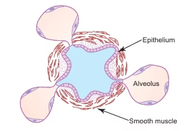
5. Alveoli
-
Alveoli form the lung parenchyma.
-
They are sac-like terminal expansions of the bronchial tree.
-
They are the site of gaseous exchange between air and blood.
-
Alveoli are compactly packed and appear honeycomb-like in sections.
-
Adjacent alveoli are separated by interalveolar septa.
-
The interalveolar septum contains capillaries, reticular and elastic fibers, and occasional fibroblasts, mast cells, and macrophages.
-
Alveolar pores of kohn may be present and help equalize air pressure.
-
Elastic fibers allow expansion during inspiration and passive recoil during expiration.
-
Reticular fibers prevent overdistension of alveoli.
Cells of alveolar lining epithelium
(a) Type i pneumocytes
-
Extremely thin squamous cells.
-
Cover about 97% of alveolar surface.
-
Contain pinocytic vesicles.
-
Form part of the blood–air barrier.
(b) Type ii pneumocytes
-
Cuboidal cells present in small groups.
-
Cover about 3% of alveolar surface.
-
Contain lamellated bodies.
-
Secrete pulmonary surfactant.
-
Surfactant reduces surface tension, prevents alveolar collapse, and has bactericidal properties.
Alveolar macrophages
-
Alveolar macrophages are derived from monocytes.
-
They form part of the mononuclear phagocytic system.
-
They phagocytose inhaled dust and carbon particles.
-
They may migrate to the alveolar surface and are expelled through sputum, which has diagnostic importance.
MCQs
1. The respiratory system is functionally divided into:
A. Upper and lower portions
B. Conducting and respiratory portions
C. Central and peripheral portions
D. Internal and external portions
Answer: B
2. The functional unit of the respiratory system is:
A. Trachea
B. Bronchus
C. Bronchiole
D. Alveolus
Answer: D
3. The conducting portion of the respiratory tract includes:
A. Alveoli
B. Alveolar ducts
C. Bronchioles
D. Trachea
Answer: D
4. The nasal cavity is divided into right and left halves by:
A. Concha
B. Turbinate
C. Nasal septum
D. Vomer
Answer: C
5. The vestibule of the nasal cavity is lined by:
A. Pseudostratified epithelium
B. Stratified squamous epithelium
C. Skin
D. Simple cuboidal epithelium
Answer: C
6. Vibrissae are present in:
A. Olfactory region
B. Respiratory region
C. Vestibule
D. Nasopharynx
Answer: C
7. Olfactory mucosa is yellow in color because of:
A. Mucus secretion
B. Lipid content
C. Lipofuscin pigment
D. Keratin
Answer: C
8. Bowman’s glands are present in:
A. Respiratory mucosa
B. Olfactory mucosa
C. Trachea
D. Bronchi
Answer: B
9. Bowman’s glands secrete:
A. Mucus
B. Serous fluid
C. Sweat
D. Surfactant
Answer: B
10. Olfactory cells are:
A. Unipolar neurons
B. Multipolar neurons
C. Bipolar neurons
D. Pseudounipolar neurons
Answer: C
11. Sustentacular cells mainly provide:
A. Sensation
B. Support
C. Secretion
D. Movement
Answer: B
12. Basal cells of olfactory epithelium are responsible for:
A. Secretion
B. Support
C. Regeneration
D. Sensation
Answer: C
13. Respiratory epithelium is:
A. Stratified squamous epithelium
B. Simple cuboidal epithelium
C. Pseudostratified ciliated columnar epithelium
D. Simple squamous epithelium
Answer: C
14. The main function of respiratory mucosa is:
A. Voice production
B. Smell
C. Conditioning of air
D. Immunity
Answer: C
15. Pharynx extends from:
A. C1 to C4
B. C2 to C7
C. Base of skull to C6
D. C3 to T1
Answer: C
16. Nasopharynx is lined by:
A. Stratified squamous epithelium
B. Simple cuboidal epithelium
C. Pseudostratified ciliated columnar epithelium
D. Simple squamous epithelium
Answer: C
17. Palatine tonsil is located in:
A. Nasopharynx
B. Oropharynx
C. Laryngopharynx
D. Trachea
Answer: B
18. Muscle coat of pharynx is composed of:
A. Smooth muscle only
B. Cardiac muscle
C. Skeletal muscle
D. Elastic fibers
Answer: C
19. The organ of voice production is:
A. Pharynx
B. Trachea
C. Larynx
D. Bronchus
Answer: C
20. Total number of laryngeal cartilages is:
A. 7
B. 8
C. 9
D. 10
Answer: C
21. Which is an elastic cartilage of larynx?
A. Thyroid
B. Cricoid
C. Arytenoid
D. Epiglottis
Answer: D
22. Vocal folds are lined by:
A. Pseudostratified epithelium
B. Stratified squamous epithelium
C. Simple squamous epithelium
D. Cuboidal epithelium
Answer: B
23. Trachea extends from:
A. C4 to T6
B. C6 to T4
C. C3 to T5
D. C5 to T3
Answer: B
24. Tracheal cartilage is:
A. Complete ring
B. Flat plate
C. C-shaped
D. Elastic plate
Answer: C
25. Trachealis muscle helps in:
A. Inspiration
B. Voice production
C. Swallowing
D. Coughing
Answer: D
26. Tracheal epithelium is:
A. Simple squamous
B. Stratified squamous
C. Pseudostratified ciliated columnar
D. Cuboidal
Answer: C
27. Intrapulmonary bronchi contain:
A. C-shaped cartilage
B. Elastic cartilage
C. Cartilage plates
D. No cartilage
Answer: C
28. Goblet cells gradually disappear in:
A. Trachea
B. Bronchi
C. Bronchioles
D. Pharynx
Answer: C
29. Bronchioles are characterized by absence of:
A. Smooth muscle
B. Elastic fibers
C. Cartilage
D. Epithelium
Answer: C
30. Clara cells are found in:
A. Trachea
B. Bronchi
C. Bronchioles
D. Alveoli
Answer: C
31. Clara cells secrete:
A. Mucus
B. Surfactant
C. Glycoprotein
D. Enzymes
Answer: C
32. Bronchoconstriction is caused by stimulation of:
A. Sympathetic nerves
B. Vagus nerve
C. Phrenic nerve
D. Intercostal nerve
Answer: B
33. Respiratory bronchioles represent:
A. Conducting zone
B. Respiratory zone
C. Transitional zone
D. Excretory zone
Answer: C
34. Alveolar ducts are lined by:
A. Cuboidal epithelium
B. Columnar epithelium
C. Squamous epithelium
D. Stratified epithelium
Answer: C
35. Lung parenchyma mainly consists of:
A. Bronchi
B. Bronchioles
C. Alveoli
D. Trachea
Answer: C
36. Alveoli give the lung its:
A. Hard texture
B. Solid structure
C. Spongy appearance
D. Fibrous nature
Answer: C
37. Interalveolar septum contains:
A. Cartilage
B. Smooth muscle
C. Capillaries
D. Serous glands
Answer: C
38. Pores of kohn help in:
A. Surfactant secretion
B. Air passage between alveoli
C. Blood circulation
D. Mucus secretion
Answer: B
39. Elastic fibers in alveoli help in:
A. Gas diffusion
B. Expansion only
C. Passive recoil
D. Surfactant formation
Answer: C
40. Type i pneumocytes cover about:
A. 50% alveolar surface
B. 70% alveolar surface
C. 90% alveolar surface
D. 97% alveolar surface
Answer: D
41. Type i pneumocytes are:
A. Cuboidal
B. Columnar
C. Squamous
D. Stratified
Answer: C
42. Type ii pneumocytes secrete:
A. Mucus
B. Glycoprotein
C. Pulmonary surfactant
D. Enzymes
Answer: C
43. Lamellated bodies are found in:
A. Type i pneumocytes
B. Macrophages
C. Type ii pneumocytes
D. Clara cells
Answer: C
44. Pulmonary surfactant function is to:
A. Increase surface tension
B. Decrease surface tension
C. Increase diffusion distance
D. Cause collapse of alveoli
Answer: B
45. Alveolar macrophages are derived from:
A. Neutrophils
B. Lymphocytes
C. Monocytes
D. Eosinophils
Answer: C
46. Alveolar macrophages are also called:
A. Foam cells
B. Dust cells
C. Kupffer cells
D. Plasma cells
Answer: B
47. Blood–air barrier is formed mainly by:
A. Type ii pneumocytes
B. Alveolar macrophages
C. Type i pneumocytes and capillary endothelium
D. Clara cells
Answer: C
48. Reticular fibers in alveoli:
A. Allow gas exchange
B. Secrete surfactant
C. Prevent overdistension
D. Cause elasticity
Answer: C
49. The respiratory epithelium cilia beat towards:
A. Alveoli
B. Lungs
C. Pharynx
D. Pleura
Answer: C
50. The main site of gaseous exchange is:
A. Bronchiole
B. Respiratory bronchiole
C. Alveolar duct
D. Alveolus
Answer: D