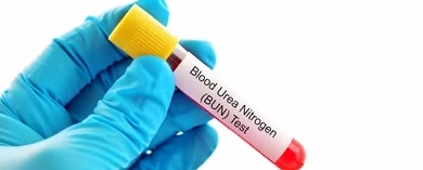
AIM: Determination of Blood urea
Non-enzymatic method
Principle of Diacetyl monoxime method
Under acidic conditions, when urea is heated with compounds containing two adjacent carbonyl groups, such as Diacetyl, in the presence of ferric ions and thiosemicarbazide, pink-coloured diazine is formed at 520 nm.
Reagent’s composition
- Sodium tungstate 10%
- Sulfuric acid 2/3 N
- Diacetyl monoxime 2% solution
- Thiosemicarbazide – 40% solution
- Sulfuric acid-phosphoric acid reagent
- Stock standard solution of urea 250 mg/100 ml
- Working standards 1 in 100 dilution
- Working reagent: Mix one part each of reagents 3 and 4 and two parts of reagent
- Prepare fresh each day.
Preparation of protein-free filtrates (PFF)
- Distilled Water 5 ml
- Blood 1 ml
- 10% Sod. Tungstate 2 ml
- 2/3 NH2SO4 2 ml
(1 ml of protein-free filtrate = 0.025 ml of blood)
Procedure
| Blank | Std. | Test | |
| Distilled water | 1 ml | – | – |
| Standard | – | 1 ml | – |
| PPF | – | – | 1 ml |
| Working Reagent | 5 ml | 5 ml | 5 ml |
Mix thoroughly and place the tubes in a boiling water bath for 30 minutes. Take the reading at 540 nm.
Enzymatic method
Principle of the urease method
Urea + H2O ——— Urease —-> 2NH3 + CO2
NH3 + α-KG + NADH ——- GLDH ——->L-Glutamate + NAD
Reagent’s composition
| R1 | |
| Tris buffer | 100 mmol/l |
| α-ketoglutarate | 5.49 mmol/l |
| Urease | ≥ 10 kU/l |
| GLDH | ≥ 3.8 kU/l |
| R2 | |
| NADH | 1.66 mmol/l |
| R3 standard |
Specimen
Use serum, EDTA plasma and heparin
Procedure
| Blank | Std. | Test | |
| Distilled water | 0.010 ml | – | – |
| Standard | – | 0.010 ml | – |
| PPF | – | – | 0.010 ml |
| Working Reagent | 1 ml | 1 ml | 1 ml |
Mix and measure the initial absorbance after 30 sec, start the timer simultaneously and read again exactly after 1 min.
Nessler’s method
NH3 + K2HgI4 ———————> Brown compound
(potassium mercuric iodide) (measured at 450nm)
Reagent’s composition
- Mixed acid reagent (MAR)
It contains orthophosphoric acid, sulphuric acid, ferric chloride and distilled water.
- Mixed colour reagent (MCR)
It contains Diacetyl monoxime and thiosemicarbazide.
Std. Conc. is 40mg/dl
Procedure
| Test | Standard | Blank | |
| Distilled water, ml | 4.0 | 3.9 | 3.9 |
| Standard (40 mg/dl), ml | – | 0.1 | – |
| PFF, ml | 0.1 | – | – |
| Mixed acid reagent, ml | 1.5 | 1.5 | 1.5 |
| Mixed coloured reagent, ml | 1.5 | 1.5 | 1.5 |
Mix thoroughly and place the tubes in a water bath for 30 minutes. Cool and read the pink-coloured solution at 520 nm.
Calculation
Conc. of blood urea = OD of test / OD of std. x Conc. of std. (40mg/dl)
BUN to urea
Mol. Wt. of urea is 60
2 atoms of N = 14 x 2 = 28
BUN mg = mg urea x
= mg urea ÷ 2.14
Normal range
Urea
20 to 40 mg/dl
BUN
- Blood urea nitrogen = 10 to 20 mg /dl
- Children (BUN) = 5 to 18 mg/dl
- Infants = 5 to 18 mg/dl
- Newborn = 3 to 12 mg/dl
Clinical significance
Azotemia: increased blood urea
Uremia: Azotemia is associated with clinical symptoms indicative of multi-organ failure, also called end-stage renal disease.
Cause of urea plasma elevation: –
- Pre-renal
- Renal
- Post-renal
Causes of increased urea (BUN)
Impaired Renal Function:
Prerenal causes
- These are mostly due to decreased blood flow to the kidneys.
- Congestive heart failure and Myocardial infarction.
- Salt and water depletion.
- Stress.
- Haemorrhage in the GI tract.
- Dehydration.
- Excessive protein catabolism.
- Burn.
Chronic Renal Diseases:
Renal causes:
- Any urinary tract obstruction also increases the BUN/creatinine ratio. In the case of protein catabolism, the serum creatinine is normal.
- Diabetes mellitus with ketoacidosis
Urinary Tract Obstruction:
Postrenal causes
- Ureteral obstruction:
- By the stone
- Cancers
- Inflammation
- Surgical procedure
- Obstruction of the bladder, urethra
- Prostatic enlargement
- Prostatic cancer
- Inflammation
- Stones
Causes of decreased Urea/BUN
- Malnutrition and a low-protein diet.
- Overhydration
- Pregnancy