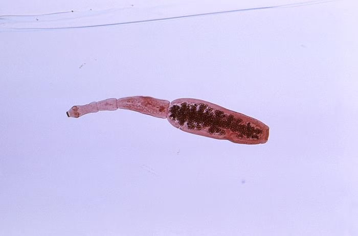
Echinococcus is a genus of parasitic tapeworms belonging to the family Taeniidae, and it causes a disease known as echinococcosis or hydatid disease. The genus includes several species, with Echinococcus granulosus (the dog tapeworm) and Echinococcus multilocularis (the fox tapeworm) being the most clinically significant to humans.
Habitat
- Echinococcus granulosus:
- The adult tapeworm resides in the small intestine of definitive hosts, primarily canids (dogs, wolves, and foxes), where it grows and releases eggs.
- The larval stage (hydatid cysts) forms in the liver and lungs and less commonly in other organs such as the bones and brain of intermediate hosts (usually herbivores like sheep, cattle, goats, and sometimes humans).
- Echinococcus multilocularis:
- The adult form lives in the intestines of foxes, dogs, and wolves.
- The larval stage forms in the liver of intermediate hosts, particularly rodents (such as voles, rats, and mice), but humans can also serve as accidental intermediate hosts.
Epidemiology
- Echinococcus granulosus (Cystic Echinococcosis or Hydatid Disease):
- Endemic in regions with large populations of dogs and livestock, particularly in South America, Europe, the Middle East, Asia, Africa, and Australia.
- Transmission occurs when definitive hosts (dogs or wild canids) ingest the hydatid cysts from infected meat of intermediate hosts (such as sheep or cattle).
- Humans become infected by ingesting Echinococcus eggs from contaminated food, water, or soil (often through close contact with infected dogs or environments).
- Echinococcus multilocularis (Alveolar Echinococcosis):
- More common in Northern Europe, North America, and Asia, where foxes or wild canids are abundant.
- Humans become accidental intermediate hosts through the ingestion of Echinococcus eggs from contaminated food, water, or direct contact with infected animals (such as dogs, foxes, or coyotes).
Morphology
- Adult Worm:
- The adult Echinococcus granulosus is a small tapeworm, measuring 3–6 mm in length, with a scolex (head) that has four suckers and a rostellum (with hooks).
- The worm has a neck and three proglottids; the last contains eggs.
- Echinococcus multilocularis is morphologically similar to E. granulosus, but typically smaller in size and with a more complex life cycle.
- Larval Stage (Hydatid Cyst):
- Echinococcus granulosus forms large, fluid-filled cysts called hydatid cysts, typically in the liver or lungs, though they can also affect other organs. These cysts contain protoscolices (larvae), which can develop into adult tapeworms once ingested by a definitive host.
- Echinococcus multilocularis forms alveolar cysts, which grow more invasively and can spread through tissues, leading to a multilocular structure that resembles a tumor.
Life Cycle
-
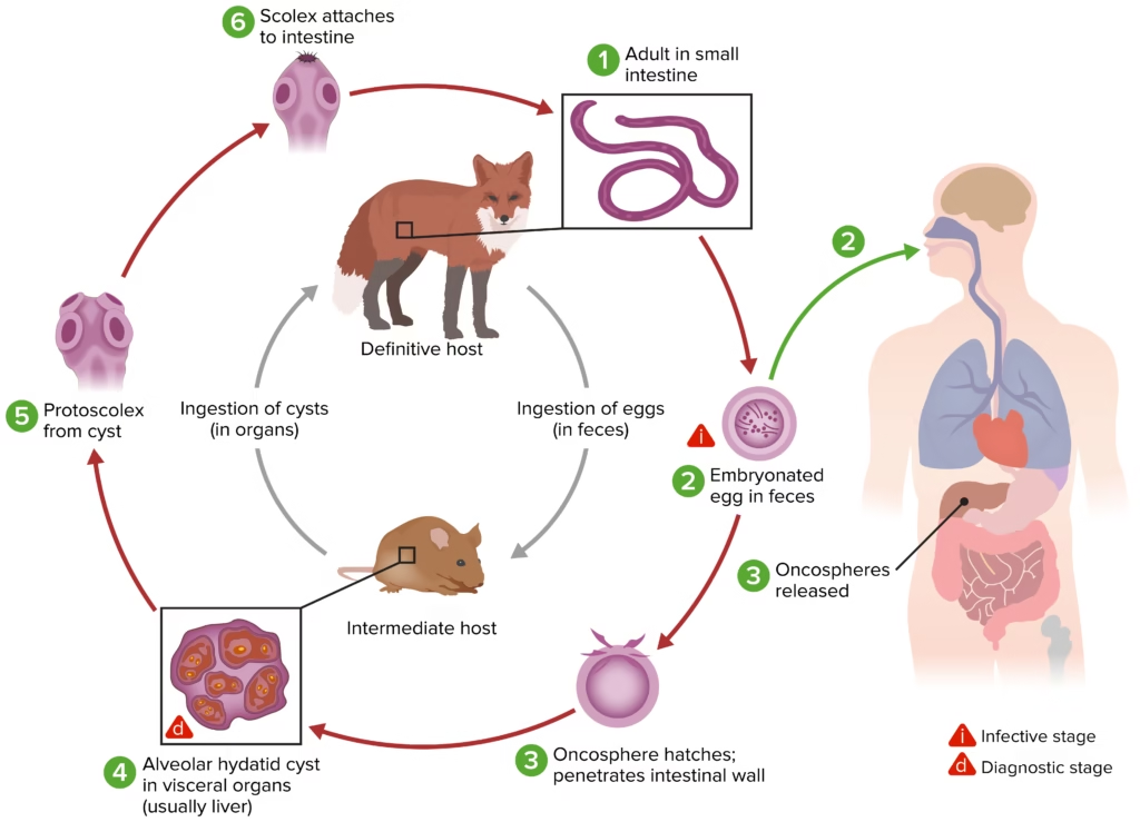
Eggs in the Environment:
- Gravid proglottids containing Echinococcus eggs are excreted by definitive hosts (canids like dogs, wolves, and foxes) into the environment, typically through feces.
- These eggs contaminate soil, water, and vegetation. Humans or intermediate hosts (herbivores, rodents) ingest the eggs by consuming contaminated food or water or contacting infected animals.
-
Larval Stage (Hydatid Cysts):
- Once ingested, the Echinococcus eggs hatch in the intestines of intermediate hosts, releasing oncospheres (larval form).
- These oncospheres penetrate the intestinal wall and migrate via the bloodstream to various organs, forming hydatid cysts in the liver, lungs, or other tissues.
- Within the cyst, the larvae develop into protoscolices.
-
Definitive Host Ingestion of Cysts:
- When the definitive host (a dog or other canid) consumes infected meat from an intermediate host, the hydatid cysts are ingested, and the protoscolices are released in the canid’s intestines.
- These protoscolices develop into adult tapeworms in the small intestine, where they mature, produce eggs, and shed them in the feces, completing the cycle.
Pathogenesis
-
Echinococcus granulosus:
- Humans are accidental intermediate hosts and become infected by ingesting Echinococcus eggs, usually through contaminated food or direct contact with infected animals.
- Once ingested, the oncospheres hatch and migrate to the liver or lungs, forming hydatid cysts.
- Over time, these cysts grow and can cause mechanical compression of surrounding organs, leading to symptoms such as abdominal pain, respiratory distress, or jaundice, depending on the cyst’s location.
- If the cyst ruptures, it can cause anaphylactic shock and spread infection to other organs.
-
Echinococcus multilocularis:
- Humans can become infected by ingesting eggs, leading to alveolar cysts (a more aggressive form of infection), primarily in the liver.
- The cysts invade surrounding tissues and may resemble a tumor, causing symptoms like abdominal pain, jaundice, and weight loss.
- Without treatment, alveolar echinococcosis can be fatal due to the invasive and tumor-like growth of the cysts.
Laboratory Diagnosis
- Microscopic Examination:
- Fecal Samples: Detection of Echinococcus eggs in fecal samples from definitive hosts (dogs, wolves, foxes) is key for diagnosing the presence of the tapeworm.
- Echinococcus granulosus eggs are round, with radial striations and an embryophore.
- Echinococcus multilocularis eggs are similar in appearance but can be distinguished by molecular tests.
- Fecal Samples: Detection of Echinococcus eggs in fecal samples from definitive hosts (dogs, wolves, foxes) is key for diagnosing the presence of the tapeworm.
- Imaging:
- Ultrasound, CT scans, or MRI visualize hydatid cysts (E. granulosus) or alveolar cysts (E. multilocularis) in the liver or other organs.
- Hydatid cysts in E. granulosus appear as well-defined, fluid-filled cysts.
- Alveolar cysts from E. multilocularis are more irregular and multiloculated, resembling tumor-like masses.
- Serological Tests:
- ELISA (Enzyme-Linked Immunosorbent Assay), indirect hemagglutination, and Western blot tests can detect Echinococcus-specific antibodies or antigens in the blood of infected individuals.
- These tests are useful for detecting Echinococcus infections in humans, especially when cysts are located in deep tissues.
- Molecular Techniques:
- Polymerase Chain Reaction (PCR) can detect Echinococcus DNA from tissue biopsies, blood, or fecal samples, accurately identifying the species involved (E. granulosus or E. multilocularis).
- PCR-RFLP (restriction fragment length polymorphism) can also differentiate between Echinococcus species.
Summary Table: Echinococcus (Hydatid Disease)
| Aspect | Echinococcus granulosus (Cystic Echinococcosis) | Echinococcus multilocularis (Alveolar Echinococcosis) |
| Habitat | Liver, lungs, and other organs of intermediate hosts (herbivores) | Liver of intermediate hosts (rodents, humans) |
| Epidemiology | Endemic in South America, Europe, Africa, Asia | Endemic in Northern Europe, North America, Asia |
| Morphology | Hydatid cysts in intermediate hosts, small adult tapeworm | Alveolar cysts in intermediate hosts, small adult tapeworm |
| Life Cycle | Involves herbivores (sheep, cattle) as intermediate hosts and canids (dogs) as definitive hosts | Involves rodents as intermediate hosts and wild canids as definitive hosts |
| Pathogenesis | Hydatid cysts in the liver/lungs leading to abdominal pain, jaundice, and rupture causing anaphylaxis | Alveolar cysts in the liver, resembling a tumor, cause jaundice, weight loss, and invasive growth |
| Laboratory Diagnosis | Microscopy (eggs in feces), serology, imaging (ultrasound/CT/MRI) | Microscopy (eggs in feces), serology, imaging (ultrasound/CT/MRI) |
Prevention and Control
- Prevention:
- Proper cooking of meat (especially liver) from intermediate hosts.
- Avoid contact with definitive hosts (dogs, foxes, and wild canids) or practice good hygiene when handling animals.
- Regular deworming of dogs to prevent them from shedding Echinococcus eggs.
- Control:
- Mass chemotherapy of humans with albendazole or mebendazole.
- Surgical removal of hydatid cysts in severe infection is often accompanied by medical treatment to prevent recurrence.