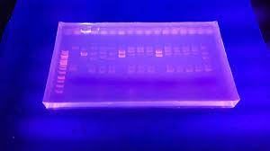
Introduction
-
Electrophoresis is a laboratory technique used to separate charged molecules such as DNA, RNA, or proteins using an electric field.
-
Molecules carry a net charge depending on their structure and the pH of the surrounding buffer, and when an electric field is applied, negatively charged molecules migrate toward the positive electrode (anode) while positively charged molecules migrate toward the negative electrode (cathode).
-
The speed of migration depends on several factors, including the molecule’s charge, size, and shape, the properties of the medium, and the strength of the electric field.
-
Agarose gel electrophoresis is commonly used for separating DNA and RNA, as it has large pores suitable for a wide range of fragment sizes.
-
Polyacrylamide gel electrophoresis (PAGE) is used for proteins and small nucleic acids, with smaller pores that allow for high-resolution separation.
-
SDS-PAGE gives proteins a uniform negative charge so that separation is based on size alone, while native PAGE preserves the molecule’s natural charge and structure.
-
Capillary electrophoresis offers high-resolution separation in narrow tubes and is widely used in DNA sequencing and forensic analysis, while isoelectric focusing separates proteins according to their isoelectric point, the pH at which they carry no net charge.
Basic Principles
-
Electric Field:
- When a voltage is applied across a medium (usually a gel or a membrane), charged particles move toward the electrode with the opposite charge. Cations move towards the cathode (negative electrode), and anions move towards the anode (positive electrode).
-
Medium:
- The separation medium is typically a gel (e.g., agarose or polyacrylamide) or a paper matrix. The medium acts as a sieve, allowing particles to migrate at different rates based on size, charge, and shape.
-
Separation Mechanism:
- Size: Smaller particles move faster through the medium, while larger particles encounter more resistance and move slower.
- Charge: Particles with greater or higher mobility move faster toward the electrode of the opposite charge.

- Shape: The shape of the particles can also affect their migration rate.
Types of Electrophoresis
-
Agarose Gel Electrophoresis:
- Medium: Agarose gel, which is a polysaccharide derived from seaweed.
- Applications: Commonly used for separating nucleic acids (DNA, RNA) based on size. It is widely used in molecular biology for DNA fragment analysis, PCR product verification, and DNA sequencing.
- Procedure: DNA samples are loaded into wells in an agarose gel, and an electric field is applied. DNA fragments migrate through the gel and are visualized using staining agents like ethidium bromide or SYBR Green.
-
Polyacrylamide Gel Electrophoresis (PAGE):
- Medium: Polyacrylamide gel provides a higher resolution than agarose gel.
- Types:
- SDS-PAGE: Separates proteins based on their size after denatured and coated with sodium dodecyl sulfate (SDS), which imparts a negative charge to the proteins.
- Native PAGE: Separates proteins based on size, charge, and shape while maintaining their native conformation.
- Applications: Used for protein analysis, including protein purification, molecular weight determination, and protein-protein interaction studies.
-
Capillary Electrophoresis (CE):
- Medium: A narrow capillary tube filled with a buffer solution.
- Applications: High-resolution separation of small molecules, peptides, proteins, and nucleic acids. It is used in clinical diagnostics, forensic analysis, and genomics.
- Procedure: A sample is introduced into a capillary tube, and an electric field is applied. Components are separated based on their charge-to-size ratio and are detected as they pass through a detection window.
-
Isoelectric Focusing (IEF):
- Medium: A gel with a pH gradient.
- Applications: Separates proteins based on their isoelectric point (pI), the pH at which a protein has no net charge. It is used for protein characterisation and analysis of protein isoforms.
- Procedure: Proteins migrate in a pH gradient until they reach the point where their net charge is zero.
-
Two-Dimensional Electrophoresis (2D-E):
- Technique: Combines IEF and SDS-PAGE.
- Applications: Provides a high-resolution separation of proteins based on their isoelectric point and molecular weight. It is used for complex protein analysis, including proteomics.
- Procedure: Proteins are first separated by IEF in one dimension and then by SDS-PAGE in the second dimension.
Procedure
1. Preparation of Gel
-
Choose agarose or polyacrylamide based on the type of sample.
-
Dissolve the gel powder in the appropriate buffer (e.g., TAE or TBE for DNA).
-
Heat until fully melted, then pour into a casting tray with a comb to form wells.
-
Allow the gel to solidify at room temperature.
2. Setting Up the Gel in the Tank
-
Place the solidified gel in the electrophoresis tank.
-
Add enough buffer to cover the gel completely.
3. Sample Preparation
-
Mix each sample with a loading dye to add density and visibility.
-
Carefully pipette the prepared samples into the wells without damaging the gel.
4. Running the Electrophoresis
-
Connect the tank to the power supply, ensuring correct electrode orientation.
-
Apply voltage and allow molecules to migrate through the gel pores.
-
Monitor progress using the tracking dye.
5. Stopping the Run
-
Turn off the power when the tracking dye approaches the end of the gel.
-
Disconnect the electrodes and remove the gel from the tank.
6. Staining the Gel
-
Immerse the gel in a suitable stain (e.g., ethidium bromide for nucleic acids, Coomassie Brilliant Blue for proteins).
-
Rinse off excess stain for clear visualization.
7. Visualisation and Documentation
-
-
Observe the separated bands under a UV transilluminator or visible light system.
-
Capture images for record-keeping and analysis.
-
Applications
-
Molecular Biology:
- DNA Fragment Analysis: Separates DNA fragments based on size for applications such as restriction fragment length polymorphism (RFLP) analysis, DNA fingerprinting, and sequencing.
- RNA Analysis: Separates RNA species for gene expression studies and RNA integrity assessment.
-
Protein Analysis:
- Protein Purification: Separates proteins from complex mixtures for further analysis or use in research.
- Protein Identification: Identifies proteins based on molecular weight and charge, often combined with mass spectrometry for detailed analysis.
-
Clinical Diagnostics:
- Haemoglobin Electrophoresis: Identifies abnormal haemoglobin variants in conditions like sickle cell disease and thalassemia.

- Protein Electrophoresis: Analyzes protein levels and types in blood samples to diagnose and monitor diseases such as multiple myeloma and liver disorders.
-
Forensic Analysis:
- DNA Profiling: Uses electrophoresis to separate and analyze DNA samples for identification and comparison in criminal investigations.
-
Biotechnology:
- Genotyping: Identifies genetic variations and mutations in research and clinical settings.
- Pharmaceutical Development: Analyzes the purity and structure of drugs and biologics.