Introduction
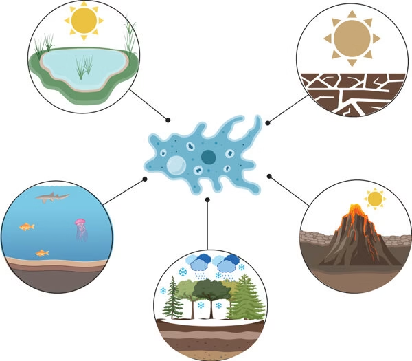
- Free-living amoebae (FLA) are protozoan parasites that thrive independently in natural environments such as soil, freshwater, air, and dust.
- These amoebae can become opportunistic human pathogens, leading to severe and often fatal infections.
- Notable genera include Acanthamoeba, Naegleria, and Balamuthia, which can cause devastating central nervous system (CNS) diseases such as granulomatous amoebic encephalitis (GAE) and primary amoebic meningoencephalitis (PAM).
- The ability of these amoebae to survive in extreme environmental conditions makes them a public health concern.
Morphology
Free-living amoebae exhibit two distinct morphological forms:
- Trophozoites:
- The active, feeding, and motile stages.
- They are characterized by an irregular shape with pseudopodia, which they use for movement and phagocytosis of bacteria, fungi, and organic debris.
- The size of trophozoites varies by species but generally ranges between 10-50 µm.
- They contain a single nucleus with a prominent central karyosome and cytoplasmic vacuoles.
- In Naegleria fowleri, trophozoites can transform into temporary flagellated forms under unfavourable conditions.
- Cysts:
- The dormant, resistant stage enables survival in harsh environmental conditions.
- Typically round or oval with a thick, protective wall composed of cellulose or other complex polysaccharides.
- Cysts are smaller than trophozoites and lack motility.
- The number of walls varies: Acanthamoeba has a double-layered cyst wall, whereas Balamuthia has a three-layered cyst wall.
- Cyst formation is triggered by unfavourable conditions such as desiccation, nutrient depletion, or chemical exposure.
Life Cycle

The life cycle of free-living amoebae consists of two primary stages:
- Trophozoite Stage:
- Under favourable conditions, amoebae exist as trophozoites, actively feeding and dividing via binary fission.
- They multiply rapidly in warm, nutrient-rich environments, thriving in stagnant water, soil, or biofilms.
- In Naegleria fowleri, trophozoites can transform into a flagellated stage when exposed to nutrient deprivation or changes in osmolarity, aiding in their dispersal.
- Cyst Stage:
- When environmental conditions become unfavourable (e.g., temperature fluctuations, drying, lack of nutrients), trophozoites encyst.
- The cysts resist extreme temperatures, chlorination, and desiccation, allowing amoebae to persist in the environment for extended periods.
- Once favourable conditions return, the cysts excyst into trophozoites, resuming their active feeding and multiplication.
- Human infections occur when trophozoites or cysts enter the body via inhalation, ingestion, or open wounds, leading to CNS and ocular infections.
Laboratory Diagnosis
Several methods are used to diagnose infections caused by free-living amoebae:
- Microscopy:
- Direct examination of clinical samples such as cerebrospinal fluid (CSF), corneal scrapings, brain tissue, or biopsy specimens.
- Wet mounts and techniques such as Giemsa, trichrome, or calcofluor white staining enhance visualization.
- Amoebic cysts and trophozoites can be identified based on size, nuclear morphology, and cytoplasmic features.
- Culture:
- Isolation using non-nutrient agar (NNA) seeded with Escherichia coli, which serves as a food source for amoebae.
- Positive cultures show amoebic tracks or growth zones around bacterial colonies, indicating trophozoite movement.
- Axenic cultures can also be maintained in specialized liquid media.
- Molecular Methods:
- Polymerase chain reaction (PCR) assays for species-specific identification of amoebic DNA in clinical samples.
- DNA sequencing and real-time PCR help differentiate closely related species.
- Fluorescent in situ hybridization (FISH) can also be used for rapid detection.
- Serology:
- Detection of anti-amoebic antibodies using enzyme-linked immunosorbent assay (ELISA) or indirect immunofluorescence.
- Serology is limited in acute infections but may help in retrospective diagnosis.
- Imaging Studies:
- Computed tomography (CT) or magnetic resonance imaging (MRI) scans can reveal brain lesions in cases of amoebic encephalitis.
- Corneal ulcerations may be observed in Acanthamoeba keratitis using slit-lamp examination.