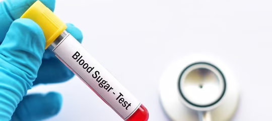
AIM: Determination of blood sugar
Introduction
- Blood sugar, also called blood glucose, is the amount of glucose (a type of sugar) present in your blood.
- Glucose is your body’s main source of energy and comes from the food you eat—especially carbohydrates like bread, rice, fruits, and sweets.
- After eating, your body breaks down these foods into glucose, which enters your bloodstream.
- A hormone called insulin, produced by your pancreas, helps move that glucose from your blood into your cells to be used for energy.
Specimen
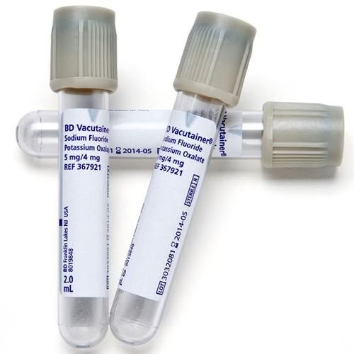
- Whole blood, plasma or serum. The container should contain a potassium oxalate-sodium fluoride mixture (3 parts potassium oxalate and one part sodium fluoride-add 3 mg/ml blood).
- Potassium oxalate prevents clotting by precipitating calcium and sodium fluoride (preventing glycolysis within the cells by inhibiting the enolase enzyme of the glycolytic pathway) for collecting whole blood and plasma.
- If serum is to be used, it should be separated immediately after clotting to avoid falsely low values of glucose concentration
Methods
- Folin-Wu method
- Ortho-toluidine method
- Glucose oxidase method
Folin-Wu Method
Principle
A protein-free filtrate is heated with an alkaline cupric tartrate solution. Glucose present in the specimen reduces cupric ions to cuprous ions, which precipitate as insoluble cuprous oxide. The amount of cuprous oxide formed is measured by reducing phosphomolybdate to molybdenum blue.
Reagents
- King’s isotonic diluent: used instead of water to minimize the interference of non-glucose-reducing substances inside the red cells.
- Sodium tungstate (10 g/dL)
- Schaffer–Hartman alkaline copper tararate
- Phosphomolybdic acid 5. Glucose standard solution (100 mg%)
Procedure
Preparation of protein-free filtrate from blood.
- In a test tube, take 7 ml of distilled water,
- 1 ml blood sample, 1 ml 10% sodium tungstate solution and 1 ml
2/3N H2SO4 solution (dropwise and with shaking). - Thus, the dilution of the blood sample is 1 in 10.
- Let it stand for 10 minutes, then filter and collect the filtrate in a dry beaker.
| Test | Standard | Blank | |
| Alkaline copper reagent, ml | 2.0 | 2.0 | 2.0 |
| PFF | 2.0 | 2.0 | 2.0 |
| Boiling water bath for 8 min. and | immediately cool | ||
| Phosphomolybdin acid, ml | 2.0 | 2.0 | 2.0 |
Read the OD at 420–490 nm or blue filter.
Calculation
Glucose concentration in mg/dl = absorbance of T/absorbance of S × 100
Orthotoluidine Method
Principle
Glucose condenses with orthotoluidine in glacial acetic acid at 100°C to form N-glucosamine, which is blue-green. Glucose concentration is proportional to the intensity of the colour
Reagents
- Orthotouidine reagent
- 10% trichloroacetic acid
- Glucose standard solution (100 mg%)
Procedure
Preparation of protein-free filtrate (PFF) from blood.
- In a dry test tube, take 3 ml of distilled water,
- 0.5 ml of blood and 1.5 ml of 10% TCA. ( Dilution of blood ≡ 1 in 10).
- Mix, keep for 10 minutes, and filter in a dry test tube to obtain a clear solution of PFF
| Test | Standard | Blank | |
| PFF, ml | 1.0 | – | – |
| Glucose standard, ml | – | 1.0 | – |
| 3% TCA, ml | – | – | 1.0 |
| Orthotoluidine reagent, ml | 5.0 | 5.0 | 5.0 |
Mix and keep the tubes in a boiling water bath for 10 min, cool and read the OD using a colourimeter with the wavelength 630–690 nm red filter.
Calculation
Glucose concentration in mg/dl = absorbance of T/absorbance of S × 100
Glucose Oxidase (GOD/POD) Method
Principle
Glucose + O2 + H2O ———> Glucose oxidase————> Gluconic acid + H2O2
2H2O2 + Phenol + 4 aminoantipyrine ——-> peroxidase ——-> Quinonimine + 4H2O
Reagents
- Phosphate buffer, pH 7.0.
- Enzyme reagent: Containing GOD, POD, 4-aminoantipyrine and phenol in phosphate buffer.
- Glucose standard: 100 mg%
Procedure
| Test | Standard | Blank | |
| Serum, ml | 0.010 | – | – |
| Glucose standard, ml | – | 0.010 | – |
| Distilled water | – | – | 0.010 |
| Enzyme reagent, ml | 1.0 | 1.0 | 1.0 |
Mix and incubate at 37°C for 15 minutes. Take OD at 530 nm and calculate the result as above.
Calculation
Plasma glucose (mg/dl) = absorbance of T/absorbance of S × 100
Normal Range
- RBS – 110 – 140 mg/dl
- FBS – 70 – 110 mg/dl
- PPBS – 110 – 140 mg/dl
Clinical significance
Hyperglycaemia
- Diabetes mellitus
- Hyperthyroidism
- Hyperpituitarism
- Increased adrenocortical activity
Hypoglycaemia
- A decrease in Blood glucose above normal. •
- Overdose of insulin for diabetic treatment.
- Hypothyroidism,
- Hypopituitarism
- Hypoadrenalism
- Insulinoma