
Introduction
- The immune system is a sophisticated defense network that protects the body from infections and diseases.
- The humoral immune response, a critical arm of the adaptive immune system, relies on B lymphocytes producing immunoglobulins (antibodies) to neutralize extracellular pathogens.
- This essay elaborates on the structure, classification, production, and functions of antibodies and the mechanisms underpinning the humoral immune response.
1. Immunoglobulins: Structure and Classification
1.1 Structure of Immunoglobulins
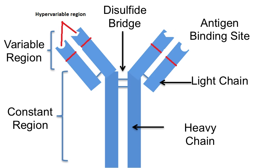
Antibodies are glycoproteins composed of four polypeptide chains: two identical heavy (H) chains and two identical light (L) chains. These chains are connected by disulfide bonds and organized into a Y-shaped structure with distinct regions: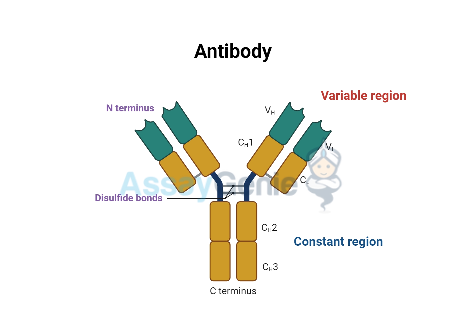
- Variable Regions (V): Found at the amino-terminal ends of both heavy and light chains, these regions are responsible for antigen binding. The variability ensures the immune system can recognize a vast array of antigens.
- The variable regions contain hypervariable or complementarity-determining regions (CDRs), which directly interact with epitopes on antigens. There are three CDRs in each variable region, maximizing diversity.
- Constant Regions (C): These regions determine the antibody’s effector functions and its classification into specific immunoglobulin isotypes (IgG, IgA, IgM, IgE, IgD).
- The constant regions interact with Fc receptors on immune cells and activate complement pathways, mediating immune defense.
- Fab Region (Fragment antigen-binding): Comprising the variable and part of the constant region, the Fab is involved in antigen recognition.
- It enables specific binding to epitopes, neutralizing pathogens or marking them for elimination.
- Fc Region (Fragment crystallizable): Composed solely of constant domains, the Fc region mediates interactions with immune cells and complement proteins.
- Different antibody classes have unique Fc regions, dictating their roles in immunity (e.g., placental transfer, allergy mediation).
- Hinge Region: Provides flexibility, allowing antibodies to bind antigens spaced variably apart.
- This flexibility enhances antigen-binding efficiency in multivalent interactions.
1.2 Classification of Immunoglobulins
The five classes of immunoglobulins are differentiated based on the structure of their heavy chains and their unique biological roles:
IgG
- Structure: Monomeric, with a gamma (γ) heavy chain.
- Function: The most abundant antibody in the serum, IgG is crucial for long-term immunity, complement activation, and opsonization. It can cross the placenta, providing passive immunity to the fetus.
- IgG subclasses (IgG1, IgG2, IgG3, IgG4) have specialized roles, such as opsonization or complement activation.
- Clinical Relevance: Elevated levels are associated with chronic infections or autoimmune diseases.
- Its long half-life (~21 days) makes it ideal for therapeutic monoclonal antibody production.
IgA
- Structure: Exists as monomers in serum and dimers in secretions (secretory IgA), stabilized by a J chain and a secretory component.
- Function: Protects mucosal surfaces by neutralizing pathogens and preventing their adherence to epithelial cells. Found in tears, saliva, and breast milk.
- Secretory IgA forms a barrier against pathogens in the respiratory, gastrointestinal, and urogenital tracts.
- Clinical Relevance: IgA deficiency is associated with recurrent mucosal infections and autoimmune disorders.
IgM
- Structure: Pentameric, with ten antigen-binding sites, held together by a J chain.
- Function: The first antibody produced during a primary immune response. Its large size makes it effective in agglutination and complement activation.
- High avidity compensates for its relatively lower affinity.
- Clinical Relevance: Indicates recent infections or early stages of immune responses.
- Elevated IgM levels may indicate acute infections or hyper-IgM syndrome.
IgE
- Structure: Monomeric, with an epsilon (ε) heavy chain.
- Function: Binds to allergens and triggers histamine release from mast cells and basophils. It also plays a role in defense against parasitic infections.
- IgE is central to Type I hypersensitivity reactions, including asthma and anaphylaxis.
- Clinical Relevance: Elevated in allergic conditions such as asthma, hay fever, and eczema.
IgD
- Structure: Monomeric, with a delta (δ) heavy chain.
- Function: Found in small amounts in the serum and primarily on the surface of immature B cells, where it acts as a receptor.
- Its exact role remains unclear but is thought to be involved in initiating B cell activation and immune tolerance.
- Clinical Relevance: Abnormal IgD levels are linked to certain cancers, such as multiple myeloma.
2. Humoral Immune Response
The humoral immune response targets extracellular pathogens by producing antibodies that neutralize or eliminate them. It is mediated by B cells, which differentiate into antibody-secreting plasma cells or memory B cells.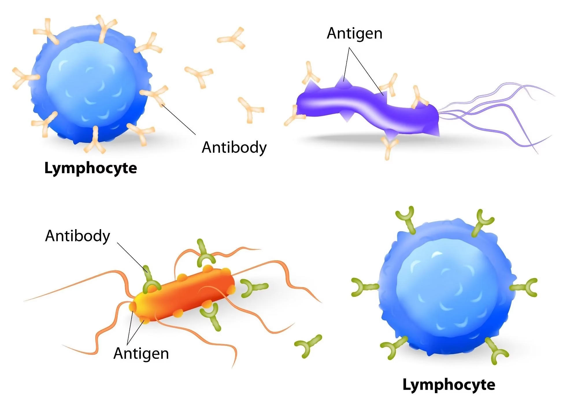
2.1 Phases of the Humoral Immune Response
Antigen Recognition
Naïve B cells possess membrane-bound immunoglobulins (IgM and IgD) that act as B cell receptors (BCRs). These receptors bind specific antigens, triggering a cascade of activation signals.
- BCRs recognize conformational or linear epitopes on antigens.
- Antigen binding induces receptor clustering and signal transduction via Igα and Igβ chains, activating B cell.
B Cell Activation
- T-Cell-Dependent Activation:
- Protein antigens are internalized by B cells, processed, and presented on MHC-II molecules.
- Helper T cells recognize the antigen-MHC complex and secrete cytokines (e.g., IL-4, IL-21) to promote B cell proliferation and differentiation.
- This process leads to germinal center formation, where affinity maturation and class switching occur.
- T-Cell-Independent Activation:
- Non-protein antigens, such as polysaccharides, directly stimulate B cells by cross-linking their receptors. This results in a short-lived response dominated by IgM production.
- Lacks the formation of memory B cells and affinity maturation.
Clonal Expansion and Differentiation
Activated B cells undergo proliferation and differentiation into:
- Plasma Cells: Short-lived cells that secrete large amounts of antibodies.
- Long-lived plasma cells can reside in the bone marrow, continuously producing antibodies.
- Memory B Cells: Long-lived cells that respond rapidly during subsequent antigen exposure.
- These cells express high-affinity BCRs due to somatic hypermutation.
2.2 Primary vs. Secondary Humoral Responses
Primary Immune Response
- Occurs upon first exposure to an antigen.
- Features a lag phase of 5–7 days before antibodies are detectable.
- IgM is the first antibody produced, followed by class switching to IgG.
- Antibody affinity is moderate due to limited somatic hypermutation.
Secondary Immune Response
- Triggered by re-exposure to the same antigen.
- Faster (1–3 days), stronger, and longer-lasting due to memory B cells.
- Characterized by high levels of IgG with increased affinity for the antigen (affinity maturation).
3. Functions of Antibodies in the Humoral Immune Response
Neutralization
Antibodies block the binding of toxins, viruses, or bacteria to host cells, preventing infection or toxic effects.
-
- For example, neutralizing antibodies against influenza virus prevent its entry into respiratory epithelial cells.
Opsonization
Antibodies coat pathogens, marking them for phagocytosis by macrophages and neutrophils.
-
- Fcγ receptors on phagocytes recognize the Fc region of IgG, enhancing pathogen uptake.
Complement Activation
IgM and IgG initiate the classical complement pathway, leading to pathogen lysis and inflammation.
-
- Complement proteins like C3b further enhance opsonization.
Agglutination
Antibodies cross-link multiple pathogens, forming clumps that are easier for immune cells to clear.
-
- IgM is particularly effective due to its pentameric structure.
Antibody-Dependent Cellular Cytotoxicity (ADCC)
IgG binds infected or abnormal cells, recruiting natural killer (NK) cells to destroy the targets via cytotoxic mechanisms.
Mucosal Defense
Secretory IgA prevents the colonization of pathogens at mucosal surfaces, safeguarding areas such as the respiratory and gastrointestinal tracts.
-
- IgA forms immune complexes that are expelled with mucus.
4. Mechanisms Underlying Antibody Diversity
The immune system generates a vast array of antibodies to recognize diverse antigens. This diversity arises through:
Somatic Recombination
Random recombination of V (variable), D (diversity), and J (joining) gene segments in heavy chains, and V/J segments in light chains, produces unique antibodies.
Junctional Diversity
Addition or deletion of nucleotides at V(D)J junctions during recombination further increases variability.
Somatic Hypermutation
Point mutations in the variable region genes enhance the affinity of antibodies for antigens during germinal center reactions.
Class Switching
B cells switch from producing IgM to other antibody isotypes (e.g., IgG, IgA) while retaining antigen specificity, tailoring the immune response to the pathogen.
5. Advantages and Limitations of Humoral Immunity
Advantages
- Specificity: Antibodies recognize unique epitopes on pathogens.
- Memory Formation: Secondary responses are faster and more robust.
- Versatility: Class switching enables adaptation to different infection types.
- Passive Immunity: Transfer of antibodies (e.g., IgG through the placenta) provides immediate protection.
Limitations
- Intracellular Pathogens: Antibodies cannot target pathogens residing within host cells.
- Antigenic Variation: Some pathogens evade detection by altering their surface antigens (e.g., influenza virus).
- Delayed Primary Response: The initial response takes several days to develop.
- Hypersensitivity Reactions: Overactive humoral responses can lead to allergies (e.g., IgE-mediated) or