
Introduction
- Microorganisms present in tissue sections are often difficult to identify with routine hematoxylin and eosin staining because many organisms are small and may resemble cellular debris.
- Special histological staining techniques are therefore used to selectively demonstrate bacteria, fungi, parasites, and other microorganisms within tissues.
- These stains work by exploiting differences in cell wall composition, chemical constituents, and staining affinity of microorganisms.
- Histological stains help localize microorganisms within tissue architecture, which is important for understanding the pattern of infection.
- Different stains are selected depending on the suspected organism, such as Gram stain for bacteria, Ziehl–Neelsen stain for acid-fast bacilli, and silver stains for fungi.
- These methods are widely used in pathology laboratories for diagnosing infectious diseases when culture results are delayed or unavailable.
- Special staining not only confirms the presence of microorganisms but also assists in differentiating between various groups of pathogens.
- Histological identification of microorganisms is clinically important because it guides accurate diagnosis, treatment, and prognosis of infectious diseases.
Gram Stain
Principle:
Gram staining is a differential staining technique used to classify bacteria into Gram-positive and Gram-negative groups based on differences in the structure of their cell wall.
- Gram-positive bacteria possess a thick peptidoglycan layer that retains the primary stain after decolorization.
- Gram-negative bacteria have a thin peptidoglycan layer and lose the primary stain during decolorization, then take up the counterstain.
The staining sequence is based on formation of a crystal violet–iodine complex:
Crystal violet + Iodine → Crystal violet-iodine complex
After alcohol treatment:
- Gram-positive bacteria retain the complex
- Gram-negative bacteria become decolorized
Materials:
- Crystal violet stain
- Gram’s iodine (mordant)
- Decolorizer (ethanol or acetone)
- Safranin or fuchsin (counterstain)
- Microscope slides
Procedure:
- Fix the tissue section on a slide by heating gently or using formalin fixation.
- Flood the slide with crystal violet and let sit for 1 minute.
- Rinse with distilled water.
- Apply Gram’s iodine for 1 minute to form the crystal violet-iodine complex.
- Rinse with water.
- Decolorize with ethanol or acetone for 10-30 seconds (or until color stops running).
- Rinse immediately with water to stop the decolorization process.
- Counterstain with safranin or fuchsin for 1 minute.
- Rinse with water and blot dry.
- Examine under a microscope. Gram-positive bacteria will appear purple, and Gram-negative bacteria will appear pink or red.
Interpretation: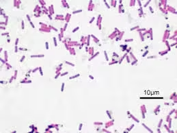
Gram-Positive Bacteria
- Appear purple or deep blue
- Retain crystal violet–iodine complex
Gram-Negative Bacteria
- Appear pink or red
- Take up counterstain after decolourization
Applications:
- Rapid preliminary identification of bacteria in clinical specimens.
- Differentiation of Gram-positive and Gram-negative organisms.
- Helps guide initial antimicrobial therapy.
- Useful in tissue sections and pus samples.
- Important for assessing specimen quality and bacterial morphology.
Acid-Fast Stain
Principle:
Acid-fast staining is a special staining technique used to identify microorganisms that possess high lipid content (mycolic acid) in their cell wall. Because of this waxy cell wall, these organisms resist decolorization by strong acid after taking up the primary stain.
The most commonly used acid-fast method is Ziehl–Neelsen stain.
In this method:
- Carbol fuchsin penetrates the waxy cell wall with the help of heat.
- Acid-fast organisms retain the red stain even after treatment with acid-alcohol.
- Non–acid-fast organisms lose the stain and take up the counterstain.
The basic reaction is:
Carbol fuchsin+Mycolic acid→Stable red staining
Materials:
- Clean glass slide
- Heat-fixed smear or tissue section
- Carbol fuchsin (primary stain)
- Acid-alcohol (decolorizer)
- Methylene blue or malachite green (counterstain)
- Distilled water
- Spirit lamp or heating source
- Microscope
Procedure:
- Fix the tissue section on a slide.
- Cover the slide with carbol fuchsin stain.
- Heat gently (do not boil) for 3-5 minutes to allow the stain to penetrate.
- Rinse with water.
- Decolorize with acid-alcohol until no color runs off (about 30 seconds).
- Rinse with water.
- Counterstain with methylene blue or malachite green for 1-2 minutes.
- Rinse with water, blot dry, and examine under a microscope. Acid-fast bacteria will appear bright pink, while other cells will appear blue or green.
Interpretation:
Acid-fast bacteria appear bright red or pink against a blue or green background.
Applications:
- Detection of acid-fast bacilli in sputum, tissue, and body fluids.
- Diagnosis of tuberculosis.
- Detection of mycobacterial infections in tissue sections.
- Identification of partially acid-fast organisms.
Silver Stain
Principle:
Silver stains are special histological staining methods used to demonstrate microorganisms that have the ability to bind or reduce silver ions. In these techniques, silver salts are deposited on microbial cell walls and then reduced to visible metallic silver, producing black or dark brown staining of organisms against a pale background.
These stains are especially useful for organisms that are difficult to visualize with routine stains, particularly fungi and certain delicate bacteria.
The reaction can be represented as:
Silver ions→Metallic silver (black deposit)
Materials:
- Tissue section or smear on glass slide
- Silver nitrate solution
- Methenamine solution
- Borax or buffer solution
- Chromic acid (oxidizing agent)
- Sodium thiosulfate
- Counterstain – light green or hematoxylin
- Distilled water
Procedure:
- Deparaffinize and hydrate tissue sections.
- Oxidize with chromic acid for 10 minutes, then rinse with water.
- Rinse in sodium bisulfite solution for 1 minute to remove excess chromic acid, then rinse with water.
- Immerse in methenamine silver solution at 60°C for 10-15 minutes.
- Rinse in distilled water.
- Tone with gold chloride solution for a few seconds.
- Rinse with water, then place in sodium thiosulfate for 1 minute to remove unreacted silver.
- Rinse and counterstain with light green if desired.
- Rinse, dehydrate, and mount for microscopic examination. Fungi will appear black, while tissue will be light green or yellowish.
Interpretation:
Positive Reaction
- Microorganisms appear black or dark brown.
Background
- The tissue background appears pale green or light-coloured, depending on the counterstain.
Characteristic Appearance
- Fungal walls stain sharply black.
Applications:
- Demonstrates fungal organisms in tissue sections.
- Detects delicate bacteria not clearly seen by routine stains.
- Useful in the diagnosis of opportunistic fungal infections.
- Helps identify organisms in tissue when routine staining is inconclusive.
Periodic Acid-Schiff Stain
Principle:
Periodic Acid–Schiff (PAS) stain is a special histological staining method used to demonstrate carbohydrate-rich structures such as glycogen, mucopolysaccharides, glycoproteins, and fungal cell walls. Many microorganisms, especially fungi, contain polysaccharides in their cell wall, which react strongly with PAS stain.
Periodic acid oxidizes adjacent glycol groups present in carbohydrates to form aldehydes. These aldehydes then react with Schiff reagent, producing a bright magenta color.
The reaction can be represented as:
Carbohydrate→Periodic acid → Aldehyde→ Schiff reagent → Magenta color
Materials:
- Tissue section or smear on glass slide
- Periodic acid solution
- Schiff reagent
- Distilled water
- Sulfurous acid rinse or running water
- Counterstain – hematoxylin or light green
- Fixative
Procedure:
- Deparaffinize and hydrate tissue sections.
- Treat with periodic acid for 5-10 minutes to oxidize carbohydrates.
- Rinse with water.
- Apply Schiff reagent for 15-30 minutes, reacting with aldehydes to form a magenta color.
- Rinse in tap water until color stabilizes (5-10 minutes).
- Counterstain with hematoxylin for 1 minute.
- Rinse, dehydrate, and mount. Fungal and carbohydrate structures will appear magenta against a blue background.
Interpretation:
Positive Reaction
- Carbohydrate-containing structures appear bright magenta or reddish-purple.
Background
- Nuclei usually stain blue with hematoxylin.
Microorganisms Demonstrated
- Fungal cell walls show strong magenta staining.
Applications:
- Demonstrates fungi in tissue sections.
- Detects polysaccharide-rich microbial cell walls.
- Useful in the diagnosis of deep fungal infections.
- Helps identify basement membranes and mucopolysaccharides in tissue pathology.
Immunohistochemical Staining
Principle:
Immunohistochemical staining (IHC) is a special staining technique based on the specific antigen–antibody reaction used to detect microbial antigens, cellular proteins, or tissue markers within tissue sections. In this method, a specific antibody binds to the target antigen present in the tissue, and the bound antibody is then visualized by a colored reaction using an enzyme-linked detection system.
The most commonly used enzymes are:
- Horseradish peroxidase (HRP)
- Alkaline phosphatase
The basic reaction is:
Antigen+Antibody→Enzyme complex→Colored reaction
Materials:
- Formalin-fixed paraffin-embedded tissue section
- Primary antibody specific to target antigen
- Secondary antibody linked with enzyme
- Buffer solution
- Blocking serum
- Chromogen (commonly DAB)
- Counterstain (hematoxylin)
- Distilled water
Procedure:
- Deparaffinize and hydrate tissue sections.
- Block non-specific binding with a blocking solution (e.g., BSA) for 10-15 minutes.
- Apply the primary antibody and incubate (often for 30-60 minutes at room temperature).
- Rinse with PBS.
- Apply the enzyme-conjugated secondary antibody and incubate.
- Rinse with PBS.
- Apply the chromogen (DAB), allowing the color to develop for 5-10 minutes.
- Rinse with water, counterstain (if desired), and mount. Targeted antigens will appear as a brown stain.
Interpretation:
Positive Reaction
- Target antigen appears as brown-colored deposit (with DAB).
Background
- Tissue nuclei appear blue after hematoxylin counterstaining.
Localization
Staining may be:
- cytoplasmic
- nuclear
- membranous
depending on antigen location.
Applications:
- Detects microorganisms directly in tissue sections.
- Identifies viral, bacterial, fungal, and parasitic antigens.
- Helps diagnose infections when routine stains are inconclusive.
- Widely used in tumor diagnosis and classification.
- Detects specific tissue markers for prognosis.
Fluorescent Staining
Fluorescent staining methods use fluorochrome-labelled dyes or antibodies to detect specific microorganisms.
Principle:
Fluorescent staining is a special staining technique in which fluorescent dyes or fluorochrome-labeled antibodies bind to specific microorganisms or cellular components and emit visible light when exposed to ultraviolet (UV) light under a fluorescence microscope.
The principle is based on the property of fluorochromes to absorb light of one wavelength and emit light of a longer wavelength, making microorganisms appear bright against a dark background.
The basic reaction can be represented as:
Fluorochrome+Target structure→Fluorescence under UV light
Materials:
- Clean glass slide with smear or tissue section
- Fluorochrome stain or fluorescent antibody
- Buffer solution
- Distilled water
- Mounting medium
- Fluorescence microscope
Procedure:
- Fix the tissue section on a slide.
- Apply auramine-rhodamine stain for 15-20 minutes.
- Rinse with water.
- Decolorize with acid-alcohol for 2-3 minutes.
- Rinse with water.
- Counterstain with potassium permanganate for 1 minute.
- Rinse, blot dry, and examine under a fluorescence microscope. Acid-fast bacteria will fluoresce yellow or orange against a dark background.
Interpretation:
Positive Reaction
- Microorganisms appear as bright fluorescent structures against a dark background.
Appearance Depends on Dye Used
- Auramine O usually gives bright yellow-green fluorescence.
- FITC gives green fluorescence.
Negative Reaction
- No fluorescence seen.
Applications:
- Rapid detection of microorganisms in clinical specimens.
- Highly useful for identifying acid-fast bacilli.
- Detects organisms when present in very small numbers.
- Useful in immunofluorescence-based diagnosis of infectious diseases.
Warthin-Starry Stain
The Warthin-Starry stain is a silver variation that detects spiral bacteria like Helicobacter pylori.
Principle:
Warthin–Starry stain is a special silver impregnation staining technique used to demonstrate delicate microorganisms that are difficult to visualize by routine staining methods. In this method, microorganisms bind silver ions, which are then reduced to visible metallic silver, producing dark black staining of organisms against a pale yellow or light brown background.
This stain is especially useful for thin spiral or curved bacteria because their delicate structure is poorly demonstrated by ordinary stains.
The basic reaction can be represented as:
Silver ions→Metallic silver (black staining of organisms) Silver
Materials:
The commonly required materials are:
- Tissue section on clean glass slide
- Silver nitrate solution
- Hydroquinone developer
- Gelatin solution
- Distilled water
- Fixative
- Water bath or controlled heating source
Procedure:
- Prepare paraffin tissue section on a clean slide.
- Deparaffinize and hydrate the section.
- Immerse the slide in silver nitrate solution.
- Add developer containing hydroquinone and gelatin.
- Incubate under controlled temperature until staining develops.
- Wash thoroughly with distilled water.
- Dry the slide and examine under microscope.
Interpretation:
Positive Reaction
- Microorganisms appear black or dark brown.
Background
- Tissue background appears pale yellow to light brown.
Characteristic Appearance
- Thin spiral organisms are sharply outlined in black.
Applications:
- Demonstrates delicate spiral and curved bacteria in tissue sections.
- Useful when routine stains fail to show microorganisms.
- Helps detect organisms in gastric biopsy and infected tissue.
Giemsa Stain
The Giemsa stain is often used in microbiology and parasitology for detecting intracellular pathogens and certain blood parasites.
Principle:
Giemsa stain is a Romanowsky-type stain composed of methylene blue, eosin, and azure dyes. It stains cellular components by combining acidic and basic dyes, allowing differentiation of nuclei, cytoplasm, and microorganisms.
- Basic dyes stain acidic cellular components such as nucleic acids blue to purple.
- Acidic dyes stain basic components pink to red.
Because many microorganisms contain nucleic acids and specific cytoplasmic structures, they take up characteristic colors and become visible within tissues or smears.
Materials:
- Clean glass slide with smear or tissue imprint
- Giemsa stain solution
- Buffered water (pH 6.8)
- Methanol (fixative)
- Distilled water
- Microscope
Procedure:
- Prepare a thin smear on a clean glass slide.
- Air dry completely.
- Fix the smear with methanol for 2–3 minutes.
- Flood the slide with diluted Giemsa stain.
- Allow staining for 15–30 minutes.
- Wash gently with buffered water.
- Air dry the slide.
- Examine under oil immersion microscope.
Interpretation:
Positive Appearance
- Nuclei stain purple-blue
- Cytoplasm stains pale blue
- Microorganisms show characteristic blue, purple, or red staining depending on species
Applications:
- Demonstrates blood parasites and intracellular microorganisms.
- Useful for detecting organisms in blood smears and tissue samples.
- Helps identify intracellular bacteria and protozoa.
Fite-Faraco Stain
The Fite-Faraco stain is a modification of the acid-fast stain that is gentler on tissue and used specifically for detecting organisms like Mycobacterium leprae.
Principle:
Fite–Faraco stain is a modified acid-fast staining technique specially designed to demonstrate weakly acid-fast organisms, particularly Mycobacterium leprae in tissue sections. Unlike routine Ziehl–Neelsen staining, this method uses mild deparaffinization and gentle decolorization so that the fragile lipid-rich cell wall of the organism is preserved.
The waxy cell wall containing mycolic acid retains carbol fuchsin, while background tissue takes up the counterstain.
The basic reaction can be represented as:
Carbol fuchsin + Mycolic acid → Persistent red staining
Materials:
- Paraffin tissue section on clean glass slide
- Xylene–peanut oil mixture for gentle deparaffinization
- Carbol fuchsin (primary stain)
- Mild acid-alcohol or sulfuric acid (decolorizer)
- Methylene blue (counterstain)
- Distilled water
Procedure:
- Prepare a paraffin tissue section on a clean slide.
- Deparaffinize gently using xylene–peanut oil mixture.
- Wash carefully and hydrate the section.
- Flood with carbol fuchsin and allow staining for an adequate time.
- Wash with water.
- Decolourise gently with a weak acid solution.
- Wash again thoroughly.
- Apply methylene blue as a counterstain.
- Wash, dry, and examine under a microscope
Interpretation:
Positive Reaction
- Acid-fast bacilli appear bright red.
Background
- Tissue appears blue.
Characteristic Appearance
- Bacilli are slender, red, and often seen in clusters within macrophages.
Applications:
- Demonstrates Mycobacterium leprae in tissue sections.
- Useful in skin biopsy diagnosis of leprosy.
- Detects weakly acid-fast organisms better than routine acid-fast methods.
- Helps assess bacterial load in tissue.