
Introduction
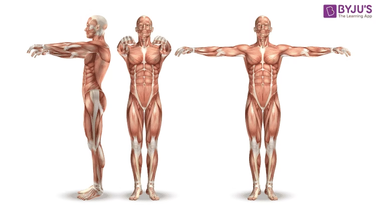
- The muscular system comprises specialized tissues capable of contracting and generating force.
- It enables movement, maintains posture, stabilizes joints, and generates heat.
- Composed of three primary muscle types (skeletal, cardiac, and smooth), it works closely with the skeletal and nervous systems to perform vital functions.
- This system allows voluntary actions, like walking and speaking, and regulates involuntary processes like heartbeat and digestion.
-
- The muscular system contributes 40-50% of total body weight.
- It plays a significant role in metabolic activities, including glucose utilization and thermogenesis.
- Muscle cells (fibers) are highly specialized and organized, ensuring efficient force generation.
Composition of the Muscular System
Muscles are composed of the following components:
Muscle Tissue
- Skeletal Muscle Tissue:
- Composed of elongated, cylindrical fibers.
- Appears striated due to the arrangement of sarcomeres.
- Cardiac Muscle Tissue:
- Made up of branched fibers with a central nucleus.
- Contains intercalated discs for synchronized contraction.
- Smooth Muscle Tissue:
- Composed of spindle-shaped cells with no visible striations.
- Found in the walls of hollow organs.
Connective Tissue
Muscles are enveloped and supported by layers of connective tissue:
- Epimysium: Surrounds the entire muscle.
- Perimysium: Covers bundles of muscle fibers called fascicles.
- Endomysium: Encloses individual muscle fibers.
Blood Supply and Innervation
- A rich blood supply ensures the delivery of oxygen and nutrients.
- Nerves control muscle contraction by transmitting signals from the central nervous system.
Proteins in Muscle Fibers
- Contractile Proteins:
- Actin (thin filament): Involved in contraction.
- Myosin (thick filament): Generates force by binding to actin.
- Regulatory Proteins:
- Troponin and Tropomyosin: Control the interaction between actin and myosin.
- Structural Proteins:
- Titin: Maintains sarcomere structure and elasticity.
- Dystrophin: Links the sarcolemma to the cytoskeleton, providing stability.
Structure of the Muscular System
Microscopic Structure
- Muscle Fiber:
- Each muscle fiber is a multinucleated cell with a sarcolemma (plasma membrane) and sarcoplasm (cytoplasm).
- Contains myofibrils, which are made up of repeating units called sarcomeres.
- Sarcomere:
- The functional unit of contraction.
- Comprised of actin and myosin arranged in overlapping patterns to create striations.
- Sarcoplasmic Reticulum (SR):
- Stores calcium ions, essential for initiating muscle contraction.
- T-Tubules:
- Allow rapid propagation of action potentials into the interior of the fiber.
Macroscopic Structure
- Muscle Belly: The thick, central part of the muscle.
- Tendons: Attach muscles to bones, enabling movement.
- Fascicles: Bundles of muscle fibers visible under the microscope.
Functions of the Muscular System
Primary Functions
- Movement:
- Skeletal muscles work with bones to produce voluntary movement.
- Posture and Stability:
- Maintains body alignment and stabilizes joints during activity.
- Heat Production:
- Muscle contractions generate heat, contributing to thermoregulation.
Additional Functions
- Circulation:
- Cardiac muscles pump blood, while smooth muscles in vessels regulate blood flow.
- Digestive Processes:
- Smooth muscles perform peristalsis to move food through the gastrointestinal tract.
- Respiration:
- Skeletal muscles like the diaphragm and intercostal muscles facilitate breathing.
- Control of Openings:
- Sphincter muscles regulate the passage of substances through hollow organs.
Types of Muscles
Muscles are categorized into three primary types based on structure and function:
Skeletal Muscle
- Appearance: Striated, multinucleated, cylindrical.
- Control: Voluntary.
- Function: Locomotion, posture, and heat production.
- Examples: Biceps brachii, quadriceps, hamstrings.
Cardiac Muscle
- Appearance: Striated, branched, single nucleus, intercalated discs.
- Control: Involuntary.
- Function: Pumps blood throughout the body.
- Location: Walls of the heart.
Smooth Muscle
- Appearance: Non-striated, spindle-shaped, single nucleus.
- Control: Involuntary.
- Function: Regulates internal movements like digestion and blood flow.
- Location: Walls of hollow organs (e.g., intestines, bladder, blood vessels).
Clinical Aspects of the Muscular System
Common Disorders
- Muscle Strain:
- Overstretching or tearing of muscle fibers.
- Muscular Dystrophy:
- The genetic condition causes progressive muscle weakness and degeneration.
- Myasthenia Gravis:
- Autoimmune disease leads to impaired communication between nerves and muscles.
- Fibromyalgia:
- Chronic disorder characterized by widespread muscle pain and fatigue.
- Rhabdomyolysis:
- Breakdown of muscle fibers, releasing myoglobin into the bloodstream, potentially causing kidney damage.
- Tendonitis:
- Inflammation of tendons due to overuse.
Diagnostic Tools
- Electromyography (EMG):
- Measures electrical activity in muscles.
- MRI and Ultrasound:
- Visualize muscle structures and detect injuries.
- Blood Tests:
- Assess levels of muscle enzymes like creatine kinase (CK) for damage.
Treatment and Management
- Physical Therapy:
- Improves muscle strength, flexibility, and recovery.
- Medications:
- Anti-inflammatory drugs for muscle pain.
- Corticosteroids for autoimmune disorders.
- Surgical Interventions:
- Tendon repair or fasciotomy for compartment syndrome.
- Lifestyle Modifications:
- Regular exercise, balanced nutrition, and adequate hydration.