Introduction
- The nervous tissue is composed of an interconnecting network of specialised cells called neurons (nerve cells) supported by
neuroglial cells. - There are about 10 million neurons in human beings.
- The function of neurons is to receive stimuli and conduct
them to a central site, the central nervous system (CNS), where they are analysed and integrated to produce a desired response
in the effector organs.
Classification
A. Morphological (based on the number of processes)
1. Unipolar neuron—has a single process (rare), e.g. mesencephalic nucleus of V cranial nerve.
2. Bipolar neuron—has two processes (an axon and a dendrite; Fig. 9.1), e.g. spiral ganglion, bipolar cells in retina, etc.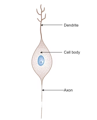
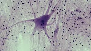
3. Multipolar neuron—has many processes (an axon and many dendrites; Fig. 9.2), e.g. autonomic ganglia motor neurons, etc.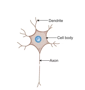
4. Pseudo-unipolar neuron—has a single process that divides into an axon (central process) and a dendrite, e.g. cranial and spinal ganglia (sensory neurons).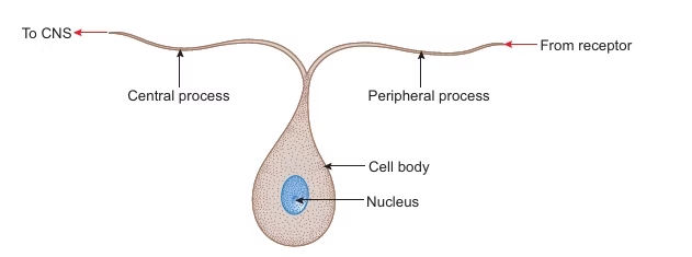
B. Functional (based on the function performed)
1. Sensory neuron—receives stimuli from receptors and conducts impulses to the CNS, e.g. sensory ganglia.
2. Motor neuron—conducts impulses from CNS to effector organs (muscles), e.g. ventral horn cells.
3. Interneuron—connects sensory and motor neurons and completes the functional circuit
Structure of Neuron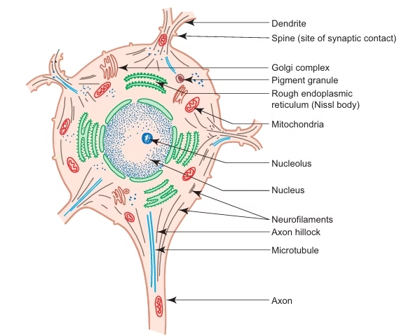
Cell body (5–150 μm):
1. Nucleus—is large, euchromatic, spherical and centrally located.
2. Nissl bodies or Nissl substance—are composed of large aggregations of rough endoplasmic reticulum– are observed as basophilic clumps by light microscopy – extend into dendrites but not into axon and axon hillock – disintegrate as a result of injury to the axon.
3. Golgi complex—is found near the nucleus.
4. Mitochondria are numerous and rod-shaped.
5. Neurofilaments (10 nm dia) and microtubules (25 nm dia)—form neuronal cytoskeleton providing structural support and intracellular transport.
6. Melanin pigments—dark brown granules.
7. Lipofuscin pigments—residual bodies not digested by lysosomes (increase with age)
Dendrites
- They are highly branched, tapering processes of a neuron. So their diameter is not uniform.
- They are covered by thorny spines (gemmules), which are sites of synaptic contact.
- Receive stimuli from sensory cells and other neurons and transmit them towards the soma.
Axon
- Single, long, cylindrical process of a neuron. So its diameter is uniform.
- Does not branch profusely, but may give rise to collaterals.
- Arises from a cone-shaped portion of the cell body called the axon hillock, which is devoid of Nissl bodies but contains bundles
of microtubules. - The cytoplasm of the axon is called axoplasm, and the plasma membrane is called axolemma.
- Terminates by dividing into many small branches, axon terminals, ending in small swellings—terminal boutons.
Myelinated and Unmyelinated Axons
- In the PNS, all axons are enveloped by Schwann cells, which provide both structural and metabolic support.
- Many axons with small diameters invaginate into one Schwann cell longitudinally and are simply surrounded by the
cytoplasm of Schwann cells. - They are called unmyelinated nerve fibres.
- In large myelinated axons, the nerve impulse jumps from node to node, resulting in faster conduction (saltatory conduction).
- The segment of myelin between two nodes of Ranvier is called an internode.
- The myelin of one internode is formed by a single Schwann cell.
Peripheral Nerve
- Each peripheral nerve (spinal or cranial) is made of bundles (fascicles) of nerve fibres (axons), which may be myelinated
and/or unmyelinated. - The bundles are held together by connective tissue, which provides structural support as well as nutritional support by
carrying blood vessels to nerve fibres.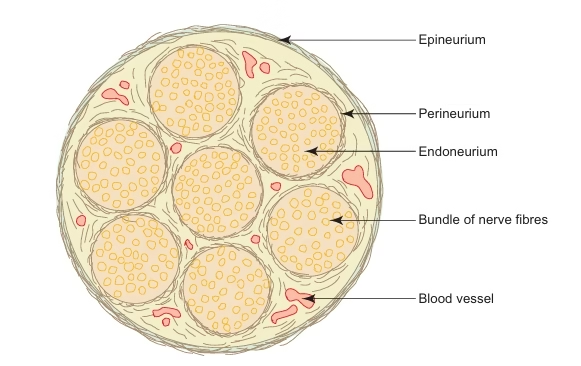
- The connective tissue framework is well appreciated in cross cross-section of a nerve, where the following structures can be observed:–
-
- Epineurium: Dense connective tissue sheath surrounding the entire nerve.
- Perineurium: A sleeve of flattened specialised epithelial cells surrounding the bundles of nerve fibres.
- Endoneurium: Loose connective tissue composed of reticular fibres supporting individual nerve fibres.
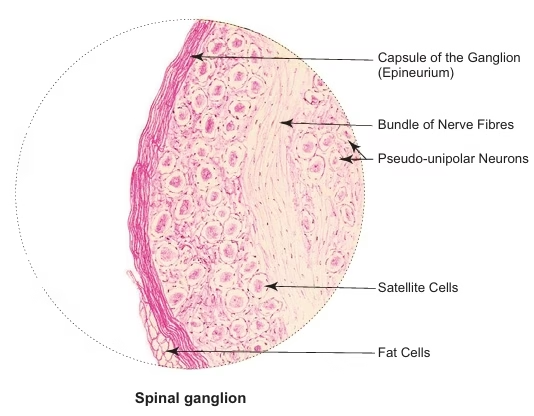
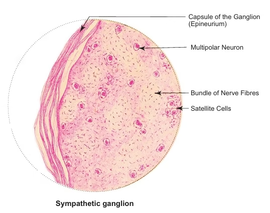
Ganglia
- Ganglia are oval bodies made of an aggregation of cell bodies of neurons outside the CNS.
- They serve as relay centres in the neuronal pathway.
- They are usually covered by a dense connective tissue capsule known as epineurium.
- The cell bodies of the neurons are enveloped by a layer of cuboidal cells called satellite cells.
- Two types of ganglia can be distinguished on the basis of morphology and function: sensory and motor ganglia.
Cerebral cortex
- The cerebral cortex consists of grey matter that covers the cerebral hemisphere.
- The surface area of the cortex is increased due to the presence of many convolutions or gyri separated by sulci.
- The cortex is made of a mixture of nerve cells, fibres, neuroglia and blood vessels.
Types of Nerve Cells
The nerve cells in the cerebral cortex are of five types. They are:
1. Pyramidal cells
-
- They are the most common type of neurons found in the cerebral cortex.
- They are pyramidal in shape.
- Their size ranges from 10 μm to 120 μm.
- Giant pyramidal cells (120 μm) in the motor cortex are called Betz cells.
- The apices of the neurons give rise to dendritic processes, which are directed towards the surface of the cortex, whereas the bases give origin to axons, which form projection fibres of the white matter.
- They are distributed in layers, 2–5, and progressively increase in size.
2. Stellate/Granule cells
-
- Small, star-shaped neurons of uniform diameter (8 μm).
- Have short axons terminating in nearby neurons.
3. Fusiform cells
-
- Spindle-shaped cells are placed at right angles to the surface in the deep layer.
- Dendrites arise from each pole of the cell body, and the axon arises from the cell body just above the lower pole and
enters the white matter.
4. Horizontal cells of Cajal
-
- They are also spindle-shaped cells, but oriented horizontally, parallel to the surface in the superficial layer.
- Dendrites arise from each pole, and an axon arises from the cell body and runs horizontally, parallel to the surface, making contact with the dendrites of pyramidal cells.
5. Cells of Martinotti: Small multipolar cells are found in layers 3–6.
-
- The axons are directed towards the surface of the cortex and generally end in the molecular layer.
Structure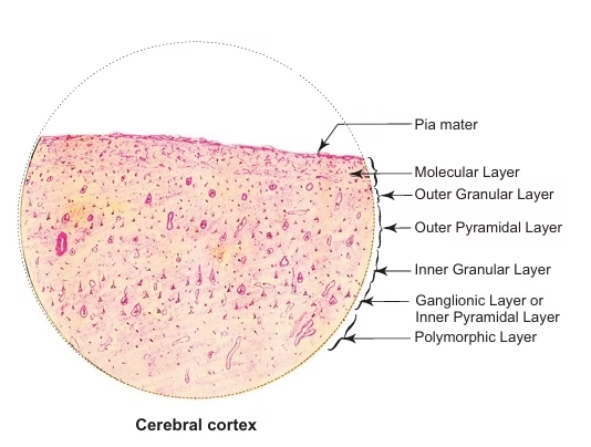
- Molecular layer (plexiform layer)—is the most superficial, well defi ned layer. It consists mainly of nerve fibres and occasional horizontal cells of Cajal.
- External granular layer—contains a large number of stellate cells and small pyramidal cells.
- External pyramidal layer—is mainly made of medium-sized pyramidal cells and also contains a few stellate cells and cells of Martinotti.
- Internal granular layer—is composed of closely packed stellate cells and a horizontally oriented white fibre band called the
outer band of Baillarger. - Internal pyramidal layer (ganglionic layer)—consists mainly of large pyramidal cells, and few stellate cells, and cells of
Martinotti. This layer also contains horizontally arranged fibres that form the inner band of Baillarger. - Multiform layer (layer of polymorphic cells)—is the deepest layer. It contains predominantly fusiform cells and also a
few stellate cells and cells of Martinotti, intermixed with many nerve fibres entering or leaving the underlying whit matter.
Cerebellar Cortex
Structure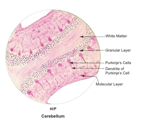
The cerebellar cortex consists of three layers: an external molecular layer, a middle Purkinje cell layer and an internal granular
layer:
1. Molecular layer
- It is the superficial thick layer and is usually lightly stained with eosin.
- Mainly made of nerve fibres and a few cells, namely, stellate cells in the superficial part and basket cells in the deeper part.
- The axons of these cells run parallel to the long axis of the folia.
- The axons of basket cells form collaterals, which arborise around the Purkinje cells in a ‘basket-like’ manner.
2. Purkinje cell layer
- Purkinje cells are large flask-shaped neurons (Golgi type I) and are arranged in a single row between molecular and granular layers.
- The dendrites of these cells pass into molecular layer and arborize profusely in a plane transverse to the folium.
- These dendrites synapse with axons of granular cells and climbing fibres that ascend to the molecular layer.
3. Granular layer
- It is stained deeply with hematoxylin because it is densely packed with very small granule neurons (the smallest cells in the body).
- The axons of these granule cells pass into the molecular layer, where they bifurcate in a T-shaped manner and run at right angles to the plane of dendritic processes of the Purkinje cells and synapse with them.
- Apart from granule cells, a few Golgi cells (type II) are also present in the granular layer. They have vesicular nuclei, more cytoplasm and short neuronal processes.
- The granule cells receive impulses from afferent Mossy fibres, which end as dilated terminals in the granular
layer.