Introduction
Paracoccidioides is a dimorphic fungus that causes paracoccidioidomycosis (PCM), a systemic fungal infection endemic to certain parts of Latin America. The disease primarily affects the lungs but can disseminate to other organs, such as the skin, mucous membranes, lymph nodes, and adrenal glands.
Key Species:
-
- Paracoccidioides brasiliensis
- Paracoccidioides lutzii
These fungi are thermally dimorphic:
-
- Environmental form: Mycelial (mold).
- Host form: Yeast.
Epidemiology
- Geographic Distribution:
-
- Endemic to South and Central America, particularly:
- Brazil, Colombia, Venezuela, Argentina, and Paraguay.
- Most cases are reported in Brazil.
- Endemic to South and Central America, particularly:
-
- Reservoir:
-
- Found in soil, especially in areas with high humidity and rich vegetation.
-
- Mode of Transmission:
-
- Inhalation of airborne conidia (infectious spores) from disturbed soil is the primary route.
- Rarely, direct inoculation into the skin can occur.
- No person-to-person transmission.
-
- At-Risk Populations:
-
- Most common in rural, agricultural workers exposed to soil.
- Male predominance: Males are 10–15 times more likely to develop the disease than females due to the protective effects of estrogen on fungal growth.
- Immunosuppressed individuals (e.g., HIV/AIDS, transplant recipients) are at higher risk of severe disease.
-
- Age Distribution:
-
- Acute/subacute form: Common in children and adolescents.
- Chronic form: Seen in adults aged 30–60 years.
-
Pathogenesis
The pathogenicity of Paracoccidioides depends on its ability to evade host defenses and adapt to the host environment.
- Inhalation of Conidia:
-
- Conidia (2–4 µm in size) are inhaled into the lungs and converted into the yeast form at 37°C.
-
- Yeast Form Adaptation:
-
- Yeasts are larger (10–30 µm) with thick, double walls, making them resistant to phagocytosis.
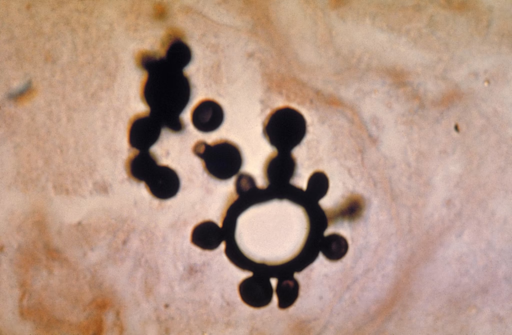
- Yeasts reproduce by multiple budding, forming the characteristic “pilot’s wheel” appearance.
-
- Immune Response:
-
- A Th1 immune response (cell-mediated) containing macrophages and CD4+ T cells is protective.
- A Th2 response (humoral immunity) or immunosuppression allows fungal dissemination.
-
- Granuloma Formation:
-
- Granulomas form in the lungs as the immune system attempts to remove the infection.
- The fungus can remain latent in granulomas for years and reactivate when immunity declines.
-
- Dissemination:
-
- If the infection is not contained, the yeast spreads hematogenously or lymphatically to other organs.
-
Clinical Manifestations
The clinical presentation of paracoccidioidomycosis varies depending on the host’s immune status and the extent of fungal spread.
- Acute/Subacute Form (Juvenile Form):
-
- Seen in children and adolescents (~10–20% of cases).
- Features:
- Rapid onset with systemic symptoms like fever, weight loss, and fatigue.
- Prominent involvement of the reticuloendothelial system:
- Generalized lymphadenopathy.
- Hepatosplenomegaly.
- Skin and mucosal lesions.
- Chronic Form (Adult Form):
-
- Most common presentation (~80–90% of cases).
- Pulmonary symptoms:
- Chronic cough, sputum production, dyspnea, and chest pain (similar to tuberculosis or COPD).
- Mucocutaneous lesions:
- Ulcerative lesions of the mouth, nose, and pharynx (“mulberry-like” appearance).
- Painful, with progressive destruction of soft tissues.
- Skin involvement:
- Papules, plaques, or nodular lesions, often on exposed areas.
- Adrenal involvement:
- It can lead to adrenal insufficiency (Addison’s disease).
- Disseminated Disease:
-
- Occurs in immunosuppressed patients.
- Involves multiple organs, including:
- CNS: Meningitis or brain abscesses.
- Bones: Osteomyelitis.
- Abdominal organs: Liver and spleen.
Laboratory Diagnosis
- Microscopy
-
- Specimens: Sputum, BAL fluid, skin scrapings, or tissue biopsies.
- Direct examination:
- Use KOH preparation or stains like Gomori methenamine silver (GMS) or Periodic acid-Schiff (PAS).
Findings:
-
- Large yeast cells (10–30 µm) with multiple buds arranged in a “pilot’s wheel” or “Mickey Mouse ears” appearance.
- Culture
-
- Specimens: Respiratory secretions, tissue biopsies, or body fluids.
- Growth conditions:
- Media: Sabouraud dextrose agar or brain heart infusion (BHI) agar.
- Dimorphic growth:
- At 25°C: Mold with septate hyphae and conidia.
- At 37°C: Yeast with characteristic multiple budding.
- Growth is slow (takes 2–4 weeks).
- Histopathology
-
- Specimens: Biopsy of infected tissues (e.g., skin, lymph nodes, or lungs).
- Findings:
- Granulomas with yeast cells showing multiple budding.
- Stains: H&E, GMS, or PAS.
- Serology
Serologic tests are highly sensitive and specific, especially in endemic areas.
Tests Available:
-
- Immunodiffusion (ID):
- Detects antibodies against Paracoccidioides antigens.
- Useful for diagnosis and monitoring treatment response.
- Complement Fixation (CF):
- Measures antibody titers.
- Higher titers indicate severe or disseminated disease.
- ELISA:
- More sensitive than ID or CF.
- Immunodiffusion (ID):
- Antigen Detection
-
- Detects Paracoccidioides antigens in blood, urine, or CSF.
- Useful for rapid diagnosis, especially in disseminated disease.
- Molecular Diagnostics (PCR)
-
- PCR-based assays detect fungal DNA in clinical specimens.
- Provides rapid and specific diagnosis.