Introduction
- The Phase Contrast Microscope was invented by Frits Zernike in 1930, for which he was awarded the Nobel Prize in 1953.
- This invention revolutionized the way scientists observe live, transparent biological specimens, which typically do not absorb light and are hard to view with traditional light microscopy.
- Before the phase contrast microscope, specimens like living cells, bacteria, and tissues were almost invisible due to their transparency.
- Zernike’s innovation allowed these specimens to be seen clearly without the need for staining, which often destroys or alters the samples.
- This non-invasive technique allows researchers to study living organisms in their natural, undisturbed state.
- The phase contrast microscope is especially beneficial in biological research, clinical diagnostics, and pharmaceutical studies where studying live, unstained cells and microorganisms is essential.
Working Principle
The phase contrast microscope works by exploiting differences in the refractive index of different parts of the sample, converting phase differences into contrast differences. Here’s a detailed breakdown of the working principle:
-
Phase Shift: As light passes through transparent specimens, such as living cells, the different parts of the sample (e.g., cell membranes, organelles) refract light to different extents. This causes a shift in the phase of the light waves.
-
Annular Diaphragm: In the condenser, an annular (ring-shaped) diaphragm is placed. This diaphragm shapes the light into a hollow cone of light that illuminates the sample. The light passing through the sample is refracted by the different refractive indices in the specimen.
-
Phase Plate: A special optical element called a phase plate is placed in the objective lens. The phase plate shifts the phase of the light that passes through the specimen by a specific amount (typically 90° or 180°). The light passing through the specimen is then recombined with the light that has passed through the rest of the system. This phase shift alters the image intensity, converting what would have been invisible (due to lack of absorption or contrast) into a visible image.
-
Interference: The phase difference between light passing through different parts of the specimen and the background is converted into an intensity difference, which enhances contrast. This process is known as interference.
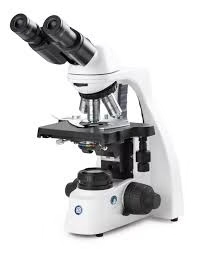
Parts of the Phase Contrast Microscope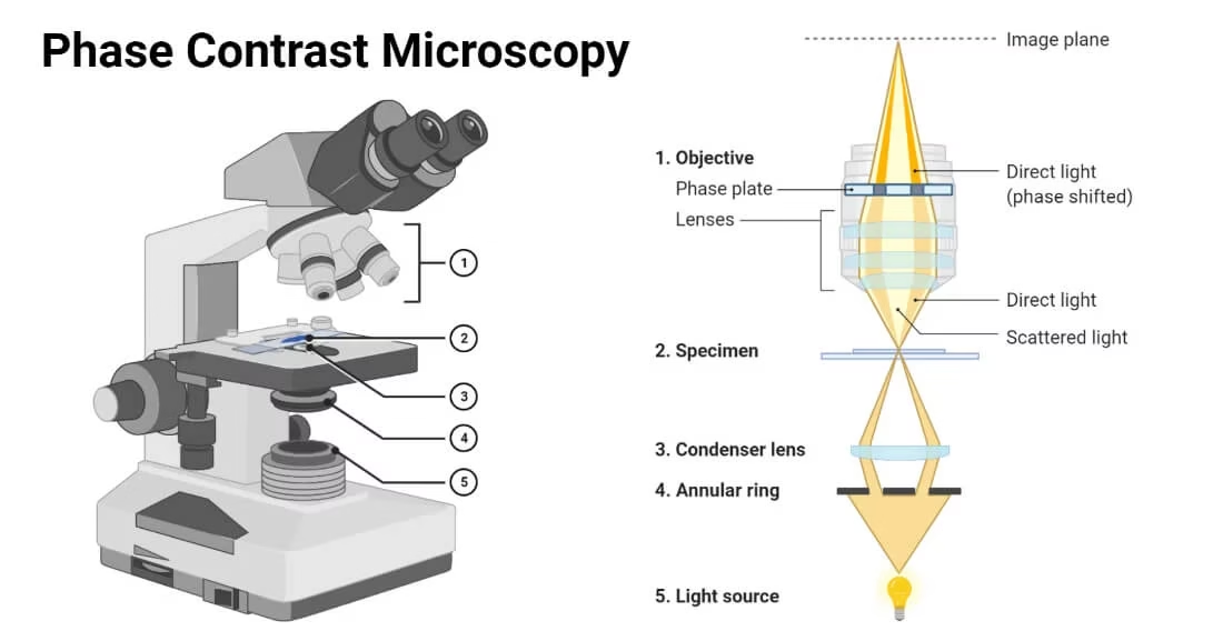
-
Light Source: Typically a bright halogen lamp or LED, the light source must provide sufficient illumination for the phase contrast to work effectively. Often, the illumination is adjustable in terms of intensity to ensure optimal lighting conditions for the sample.
-
Condenser System: The phase contrast microscope uses a specialized condenser with an annular diaphragm. The diaphragm controls the light to pass through the specimen in the form of a hollow cone, which is key for creating phase shifts.
-
Objective Lenses: These lenses are equipped with phase rings that interact with the light passing through the sample, introducing phase shifts in the light to enhance contrast. The objective lenses are often labeled with a phase contrast symbol to indicate their compatibility.
-
Phase Plate: The phase plate sits in the optical path and introduces a phase shift in the light after it passes through the sample. The phase plate modifies the light in a way that enhances the contrast of the structures within the sample.
-
Eyepiece (Ocular Lens): Standard eyepieces are used for viewing the magnified image, but with the added benefit of enhanced contrast due to the phase contrast technique.
-
Mechanical Stage: The stage allows precise movement of the specimen on the stage to ensure that different areas of the sample can be viewed.
Maintenance of the Phase Contrast Microscope
Regular maintenance of the phase contrast microscope ensures accurate results and longevity. Here’s a more detailed look at the maintenance procedures:
-
Optical Alignment: The phase rings, phase plate, and condenser system should be regularly checked for correct alignment. Misalignment can lead to poor contrast or distorted images.
-
Cleaning: Keep the lenses and optical components clean by gently wiping them with appropriate cleaning materials. Dust and fingerprints can distort the image quality.
-
Checking the Light Source: The brightness and consistency of the light source should be checked regularly. Dim or uneven light can affect the phase contrast, making it difficult to distinguish between the different structures in the sample.
-
Calibrating the Condenser: The condenser should be checked to ensure that the annular diaphragm is in the proper position for optimal illumination of the sample. This step is important for maintaining sharp contrast in the final image.
-
Ensuring Proper Temperature Control: Many phase contrast microscopes, especially in biological labs, are used to view living organisms. Keeping the temperature regulated in the microscope environment is essential to maintain live cell integrity.
Applications of Phase Contrast Microscope
The phase contrast microscope has a wide range of applications across various fields:
-
Cell Biology: It allows for studying cell division (mitosis and meiosis), cell structure, organelle function, and intracellular dynamics in living cells without using invasive stains.
-
Microbiology: The technique is indispensable for studying bacteria, yeast, and other microorganisms. It enables scientists to observe the morphology and behavior of these organisms in their natural state.
-
Medical Diagnostics: It is used in clinical laboratories to study human cells, including blood cells and tissue cultures, providing valuable information without the need for staining or fixation.
-
Neurobiology: Phase contrast microscopy is used to study neural cells, axons, dendrites, and synaptic structures in live tissue.
-
Pharmaceuticals: Used to monitor the effects of drugs on live cells, such as observing cytotoxicity, drug penetration, or cellular responses to therapeutic agents.
-
Embryology: Phase contrast is used to study early-stage embryos, cell differentiation, and growth without disturbing the specimen.
Advantages of Phase Contrast Microscope
-
No Need for Staining: Since the technique enhances contrast without altering or staining the sample, it’s ideal for studying live cells or organisms, preserving their natural structure.
-
Higher Contrast: The microscope provides significantly higher contrast compared to brightfield microscopy, making transparent structures like cell membranes, nuclei, and organelles visible.
-
Non-Destructive: Because the technique doesn’t require staining or fixation, the specimen remains unharmed and can be studied over time in its natural state.
-
Simple Setup: The phase contrast microscope is relatively easy to use once calibrated, making it accessible for routine use in laboratories.
Disadvantages of Phase Contrast Microscope
-
Halo Effect: The enhancement of contrast can result in halo effects or artificial outlines around structures, which may obscure fine details, especially in thicker samples.
-
Lower Resolution: While the contrast is significantly improved, the resolution is not enhanced, and fine details at a very high magnification may still be challenging to resolve.
-
Limited Depth of Field: The contrast enhancement can reduce the depth of field, which may make it challenging to view the entirety of thick or three-dimensional specimens.
-
Artifacts: Incorrect alignment or issues with the phase plate may cause optical artifacts, leading to images that are difficult to interpret or misleading.
Limitations of Phase Contrast Microscope
-
Not Effective for Opaque Samples: The technique is most effective for transparent specimens, and does not perform well with opaque or highly pigmented samples.
-
Requires Skilled Operators: Phase contrast microscopes require proper alignment and calibration of the optical components, meaning that users must be skilled to prevent artifacts and ensure quality imaging.
-
Optical Artifacts: Because the phase contrast microscope amplifies phase differences, some artifacts (e.g., halos or false contrast) may appear, particularly in samples with uneven thickness or refractive properties.
-
Cost and Complexity: These microscopes tend to be more expensive than standard brightfield microscopes, and they require careful setup and maintenance.