Introduction
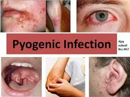
- Pyogenic infections, characterized by pus formation, are primarily caused by pathogenic bacteria that induce acute inflammation.
- These infections can manifest in various forms, including abscesses, cellulitis, pneumonia, and osteomyelitis.
- Laboratory diagnosis is critical for identifying the causative organisms, guiding treatment, and managing public health concerns.
- This detailed overview will cover the common bacterial pathogens associated with pyogenic infections, methods for sample collection, laboratory techniques for diagnosis, interpretation of results, and specific case studies.
Common Bacterial Pathogens in Pyogenic Infections
- Staphylococcus aureus
- Characteristics: Gram-positive cocci, often in clusters, catalase-positive, coagulase-positive.
- Associated Infections: Skin infections (furuncles, carbuncles), osteomyelitis, pneumonia, endocarditis, and sepsis.
- Streptococcus pyogenes (Group A Streptococcus)
- Characteristics: Gram-positive cocci in chains, beta-hemolytic, bacitracin-sensitive.
- Associated Infections: Pharyngitis, impetigo, cellulitis, and necrotizing fasciitis.
- Escherichia coli
- Characteristics: Gram-negative rods, lactose fermenter, oxidase-negative.
- Associated Infections: Urinary tract infections (UTIs), intra-abdominal infections, and neonatal sepsis.
- Klebsiella pneumoniae
- Characteristics: Gram-negative rods and lactose fermenters produce a thick capsule.
- Associated Infections: Pneumonia, UTIs, and liver abscesses.
- Pseudomonas aeruginosa
- Characteristics: Gram-negative rods, non-lactose fermenter, oxidase-positive, known for antibiotic resistance.
- Associated Infections: Respiratory infections in cystic fibrosis patients, wound infections, and bacteremia.
Sample Collection
Sample collection is crucial for accurate diagnosis. The type of sample collected depends on the site of infection:
- Skin and Soft Tissue Infections
- Abscesses: Aspiration with a sterile needle or incision and drainage for pus samples.
- Cellulitis: Swabs from the inflamed area; however, deep tissue samples yield more reliable results.
- Respiratory Tract Infections
- Sputum: Collected via expectoration; quality is assessed (e.g., leukocyte count) to ensure an adequate sample.
- Pleural Fluid: Collected via thoracentesis for suspected empyema and sent for culture and cytology.
- Urinary Tract Infections
- Urine: A clean-catch midstream sample is preferred to minimize contamination; catheterized samples may be used in certain cases.
- Bone and Joint Infections
- Synovial Fluid: Collected via arthrocentesis for suspected septic arthritis; analyzed for white blood cell counts and culture.
- Bone Biopsy: In cases of osteomyelitis, a bone sample is taken from the infected site.
- Bloodstream Infections
- Blood Cultures: Drawn in sterile conditions, typically in pairs from different sites (e.g., peripheral veins) to enhance yield.
Laboratory Techniques for Diagnosis
Culture Methods
A. Bacterial Culture:
- Media Selection: The choice of cultural media is critical. Common media include:
- Blood Agar: Supports a wide range of bacteria and helps identify hemolytic activity.
- MacConkey Agar: Selective for Gram-negative bacteria, differentiating lactose fermenters from non-fermenters.
- Chocolate Agar: Used for fastidious organisms, particularly in respiratory infections.
- Incubation Conditions: Cultures are typically incubated at 35-37°C. Anaerobic conditions may be necessary for certain pathogens.
B. Identification of Isolates:
- Colony Morphology: Observing characteristics such as size, shape, color, and hemolytic patterns helps in preliminary identification.
- Gram Staining: This fundamental technique helps distinguish between Gram-positive and Gram-negative bacteria.
Biochemical Tests
- Catalase Test: Differentiates between Staphylococcus (catalase-positive) and Streptococcus (catalase-negative).
- Coagulase Test: Identifies Staphylococcus aureus (coagulase-positive) from coagulase-negative staphylococci.
- Oxidase Test: Used to identify Pseudomonas aeruginosa (oxidase-positive) and differentiate it from Enterobacteriaceae.
- Lactose Fermentation: Observed on MacConkey agar to differentiate E. coli and Klebsiella pneumoniae from other Gram-negative bacteria.
Molecular Methods
A. Polymerase Chain Reaction (PCR):
- Nucleic Acid Amplification: PCR allows for rapid identification of specific pathogens, particularly in cases where cultures may be negative.
- Real-Time PCR: This technique quantifies bacterial DNA, providing insights into bacterial load and facilitating diagnosis of mixed infections.
B. Nucleic Acid Hybridization:
- Fluorescent In Situ Hybridization (FISH): This method can identify specific bacteria in clinical samples using fluorescently labeled probes, useful in complex infections.
Serological Tests
- Antigen Detection: ELISA and rapid antigen tests can identify bacterial antigens in samples (e.g., throat swabs for Streptococcus pyogenes).
- Antibody Detection: Serological tests may indicate past exposure or infection but are less reliable for acute pyogenic infections.
Imaging Studies
Imaging can assist in the diagnosis of pyogenic infections:
- Ultrasound: Helpful for identifying abscesses or fluid collections.
- CT Scans: Useful in evaluating deep-seated infections such as osteomyelitis or intra-abdominal abscesses and can guide drainage procedures.
Interpretation of Results
Culture Results
- Positive Culture: The growth of a pathogen confirms infection; identification helps tailor treatment.
- Negative Culture: A negative result may indicate:
- Infection by fastidious organisms (e.g., certain Streptococcus species).
- Recent antibiotic use affecting culture results.
- Contamination or inadequate sampling.
Sensitivity Testing
Antibiotic susceptibility testing (AST) is critical for guiding treatment:
- Disk Diffusion Method (Kirby-Bauer): Measures the diameter of inhibition zones around antibiotic disks to classify susceptibility.
- Minimum Inhibitory Concentration (MIC): Determines the lowest concentration of an antibiotic that inhibits bacterial growth, providing precise guidance for treatment.
Interpretation of Sensitivity Results
- Susceptible: Indicates that the antibiotic is likely effective for treating the infection.
- Intermediate: Indicates uncertain efficacy; higher doses or alternative therapies may be necessary.
- Resistant: Indicates that the pathogen is unlikely to respond to the antibiotic, necessitating alternative treatments.
Common Pyogenic Infections and Their Laboratory Diagnosis
-
Skin and Soft Tissue Infections
Pathogens: Primarily Staphylococcus aureus and Streptococcus pyogenes.
Diagnostic Methods:
-
- Culture of Abscess Pus: Direct culture from the site provides the best yield.
- Swab from Wound or Lesion: Useful but less reliable than aspirates.
Tests: Gram stain, coagulase test, and antibiotic susceptibility testing.
-
Pneumonia
Pathogens: Commonly caused by Streptococcus pneumoniae, Staphylococcus aureus, and Klebsiella pneumoniae.
Diagnostic Methods:
-
- Sputum Culture: Requires good-quality sputum; a Gram stain is performed first to assess the sample.
- Blood Cultures: Important in severe cases or when pneumonia is associated with bacteremia.
Tests: Gram stain, biochemical tests for identification, and susceptibility testing.
-
Urinary Tract Infections (UTIs)
Pathogens: Mainly Escherichia coli and Klebsiella pneumoniae.
Diagnostic Methods:
-
- Urine Culture: A midstream clean-catch urine sample is collected and cultured.
- Rapid Tests, Such as dipstick urinalysis for leukocyte esterase and nitrite.
Tests: Identification via biochemical tests and susceptibility testing.
-
-
Osteomyelitis
-
Pathogens: Frequently caused by Staphylococcus aureus and occasionally by Gram-negative rods.
Diagnostic Methods:
-
- Bone Biopsy: Preferred method for culture to confirm the pathogen.
- Imaging: X-rays, MRI, or CT scans to assess the extent of infection and detect abscesses.
Tests: Gram stain, culture, and susceptibility testing.
-
-
Bloodstream Infections
-
Pathogens: Staphylococcus aureus and Streptococcus pneumoniae are among the common culprits.
Diagnostic Methods:
-
- Blood Cultures: Drawn from two separate sites to enhance the detection of pathogens.
- Timing: Multiple cultures may be drawn over 24 hours if the initial results are negative but clinical suspicion remains high.
Tests: Identification and susceptibility testing following culture growth.