Introduction
- Bone is a specialised form of connective tissue that provides structural support to the body.
- Approximately one-third of bone consists of organic matrix, mainly collagen fibers.
- Approximately two-thirds of bone consists of inorganic mineral salts, predominantly calcium phosphate, with smaller amounts of calcium carbonate and trace elements.
- The inorganic mineral component imparts hardness and rigidity to bone.

- This hardness enables bone to withstand compressive forces during weight-bearing and impact activities.
- The organic collagen matrix provides toughness and elasticity to bone.
- This organic component allows bone to resist tensile and bending forces.
- Despite its hardness, bone is a living and highly vascular tissue with continuous metabolic activity.
- Bone undergoes constant remodelling, growth, and repair throughout life.
- Bone has a remarkable regenerative capacity and adapts structurally to mechanical stress, showing atrophy with disuse and hypertrophy with increased use.

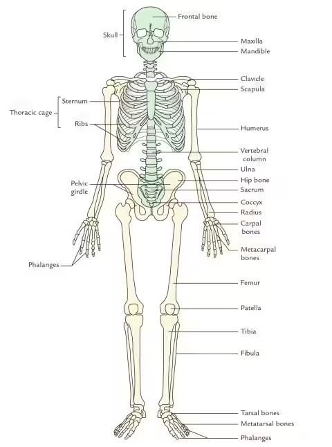
Divisions of the Skeletal System
| Division | Region / Part | Bones Included | Number of Bones |
|---|---|---|---|
| Axial Skeleton | Skull | Cranium | 8 |
| Facial bones | 14 | ||
| Hyoid bone | 1 | ||
| Auditory ossicles (Malleus, Incus, Stapes) | 6 | ||
| Vertebral Column | Cervical, thoracic, lumbar vertebrae, sacrum, coccyx | 26 | |
| Thorax | Sternum | 1 | |
| Ribs | 24 | ||
| Appendicular Skeleton | Pectoral (Shoulder) Girdle | Clavicle | 2 |
| Scapula | 2 | ||
| Upper Extremity | Humerus | 2 | |
| Radius | 2 | ||
| Ulna | 2 | ||
| Carpals | 16 | ||
| Metacarpals | 10 | ||
| Phalanges | 28 | ||
| Pelvic (Hip) Girdle | Hip bones | 2 | |
| Lower Extremity | Femur | 2 | |
| Patella | 2 | ||
| Tibia | 2 | ||
| Fibula | 2 | ||
| Tarsals | 14 | ||
| Metatarsals | 10 | ||
| Phalanges | 28 |
| Bone | Number |
|---|---|
| Frontal | 1 |
| Parietal | 2 |
| Temporal | 2 |
| Occipital | 1 |
| Sphenoid | 1 |
| Ethmoid | 1 |
| Bone | Number |
|---|---|
| Maxilla | 2 |
| Palatine | 2 |
| Zygomatic | 2 |
| Nasal | 2 |
| Lacrimal | 2 |
| Inferior nasal concha | 2 |
| Vomer | 1 |
| Mandible | 1 |
Total Number of Bones = 206
Functions
- Support – The skeleton forms the structural framework of the body and supports soft tissues.
- Protection – It protects vital organs such as the brain, spinal cord, heart, and lungs.
- Movement – Bones act as levers and provide attachment sites for muscles, enabling body movement.
- Mineral Storage – Bones store essential minerals, especially calcium and phosphorus.
- Blood Cell Formation (Hematopoiesis) – Red bone marrow produces red blood cells, white blood cells, and platelets.
- Fat Storage – Yellow bone marrow stores fat as an energy reserve.
- Maintenance of Body Shape – The skeleton gives the body its characteristic shape and posture.
- Transmission of Sound – Auditory ossicles in the middle ear help in hearing by transmitting sound vibrations.
- Acid–Base Balance – Bone helps maintain acid–base balance by buffering blood pH.
- Adaptation to Mechanical Stress – Bone remodels itself according to physical stress and strain.
Classification of Bones
A. According to Shape
1. Long Bones
- Long bones are those in which length is greater than breadth and thickness.
- They consist of a shaft called diaphysis and two expanded ends called epiphyses.
- The shaft encloses a medullary cavity containing bone marrow.
- The outer part is made of compact bone, while the ends contain spongy bone.
- Long bones act as levers for movement and support body weight.
- They are mainly present in the limbs.
- Examples: Femur, humerus, radius, ulna, tibia, fibula, metacarpals, metatarsals, phalanges.
2. Short Bones
- Short bones are approximately equal in length, breadth, and thickness.
- They are usually cuboidal in shape.
- The outer surface is made of a thin layer of compact bone, while the interior contains spongy bone.
- They provide strength and stability with limited movement.
- These bones are commonly found in regions where strength is required with flexibility.
- Examples: Carpals of wrist and tarsals of ankle.
3. Flat Bones
- Flat bones are thin, flattened, and often curved.
- They consist of two layers of compact bone enclosing spongy bone in between.
- In skull bones, the spongy layer is called diploë.
- Their main function is protection of vital organs and providing broad surface for muscle attachment.
- Examples: Skull bones, sternum, ribs, scapula.
4. Irregular Bones
- Irregular bones have complex shapes that do not fit into other categories.
- Their structure varies according to function.
- They contain a thin outer layer of compact bone with internal spongy bone.
- They provide specialized support and protection.
- Examples: Vertebrae, hip bone, sphenoid, ethmoid, mandible.
5. Sesamoid Bones
- Sesamoid bones are small round bones embedded within tendons.
- They develop in places where tendons pass over joints and experience friction.
- Their function is to reduce friction, modify pressure, and improve mechanical efficiency of muscles.
- They protect tendons from excessive wear.
- Examples: Patella (largest sesamoid bone), pisiform.
6. Pneumatic Bones
- Pneumatic bones contain air-filled cavities or sinuses lined by mucous membrane.
- These air spaces make bones lighter in weight.
- They also help in resonance of voice and conditioning of inspired air.
- Most are present in the skull.
- Examples: Frontal bone, ethmoid bone, sphenoid bone, maxilla, temporal bone.
7. Sutural (Wormian) Bones
- Sutural bones are small accessory bones present within cranial sutures.
- They are irregular in size and number.
- They develop from additional ossification centers.
- They are commonly seen in the lambdoid suture.
- Their presence may vary among individuals.
B. Developmental Classification
Developmental classification is based on the mode of ossification, that is, the method by which bones develop during embryonic life.
a. Membranous Bones
- Membranous bones develop directly from mesenchymal connective tissue membrane without a cartilage precursor.
- The process of formation is called intramembranous ossification.
- Mesenchymal cells differentiate into osteoblasts, which begin secreting bone matrix.
- These bones appear early during fetal life and mainly form protective flat bones.
- They are usually associated with protection of delicate organs such as the brain.
- Examples: Frontal bone, parietal bone, nasal bone, maxilla, most of mandible.
b. Cartilaginous Bones
- Cartilaginous bones develop from a pre-existing hyaline cartilage model.
- The process of formation is called endochondral ossification.
- First, cartilage is formed, then gradually replaced by bone tissue.
- This type of ossification is responsible for formation of most long bones and many irregular bones.
- Growth in length of long bones occurs through epiphyseal cartilage until maturity.
- Examples: Femur, humerus, tibia, fibula, vertebrae, ribs.
c. Membranocartilaginous Bones
- These bones develop partly by intramembranous ossification and partly by endochondral ossification.
- One part of the bone is formed directly in membrane, while another part develops from cartilage.
- They represent mixed developmental origin.
- Examples: Clavicle, occipital bone, temporal bone, mandible.
C. Regional Classification
Regional classification is based on the location of bones in the body.
a. Axial Skeleton
- Axial skeleton forms the central longitudinal axis of the body.
- It supports the head, neck, and trunk.
- Its main function is protection of vital organs and maintenance of posture.
- It includes:
- Skull
- Vertebral column
- Ribs
- Sternum
- Total number of bones = 80
Components of Axial Skeleton
- Skull = 22 bones
- Auditory ossicles = 6 bones
- Hyoid bone = 1 bone
- Vertebral column = 26 bones
- Sternum = 1 bone
- Ribs = 24 bones
b. Appendicular Skeleton
- Appendicular skeleton includes bones of limbs and girdles attached to axial skeleton.
- It is mainly concerned with movement and locomotion.
- It includes:
- Pectoral girdle
- Upper limbs
- Pelvic girdle
- Lower limbs
- Total number of bones = 126
Components of Appendicular Skeleton
- Pectoral girdle = 4 bones
- Upper limbs = 60 bones
- Pelvic girdle = 2 bones
- Lower limbs = 60 bones
D. Structural Classification
Structural classification is based on gross appearance and internal arrangement of bone tissue.
a. Compact Bone (Cortical Bone)
- Compact bone is dense, hard, and solid.
- It forms the outer shell of all bones.
- It consists of closely packed Haversian systems (osteons).
- Each osteon contains:
- Central Haversian canal
- Concentric lamellae
- Lacunae with osteocytes
- Canaliculi
- It provides mechanical strength and resistance to bending and weight-bearing forces.
- It is abundant in shaft of long bones.
b. Cancellous Bone (Spongy Bone / Trabecular Bone)
- Cancellous bone consists of a network of bony trabeculae.
- Spaces between trabeculae contain bone marrow.
- It is lighter than compact bone.
- It is metabolically more active than compact bone.
- It helps absorb shock and distribute forces.
- Found mainly in:
- Epiphysis of long bones
- Interior of flat bones
- Vertebrae
c. Lamellar Bone
- Lamellar bone is mature bone in which collagen fibers are arranged in regular lamellae.
- It has organized microscopic architecture.
- It is mechanically strong and stable.
- Present in normal adult skeleton.
d. Woven Bone
- Woven bone is immature bone with irregular arrangement of collagen fibers.
- It is formed rapidly during:
- Fetal life
- Fracture healing
- Certain pathological conditions
- It is weaker than lamellar bone.
- Later it is replaced by lamellar bone.
Blood Supply of Bones
- Bone is a highly vascular tissue and receives rich blood supply essential for growth, nutrition, and repair.
- Blood vessels enter bone through nutrient foramina present on the surface of bones.
- The main artery supplying a long bone is called the nutrient artery.
- Nutrient artery enters the shaft through the nutrient foramen and passes obliquely through the nutrient canal.
- Inside the medullary cavity, the nutrient artery divides into ascending and descending branches to supply:
- Bone marrow
- Endosteum
- Inner two-thirds of cortex
- Periosteal arteries arise from surrounding blood vessels and supply the periosteum and outer one-third of cortical bone.
- Metaphyseal arteries supply the metaphysis and arise from neighboring systemic vessels.
- Epiphyseal arteries supply the epiphysis and are especially important during growth.
- In growing bones, epiphyseal and metaphyseal vessels are separated by the epiphyseal plate, but after fusion they communicate freely.
- Venous drainage of bone follows arteries through nutrient veins and periosteal veins.
- Blood supply is very important for fracture healing because osteogenic cells and nutrients reach the injured area through blood vessels.
- Interruption of the blood supply may lead to avascular necrosis of bone.
Nerve Supply of Bones
- Bones receive a rich nerve supply, especially through the periosteum.
- Nerves enter bone along with blood vessels through the nutrient foramen.
- The periosteum is highly sensitive because it contains numerous sensory nerve endings.
- These sensory nerves are mainly pain fibers, making periosteum very sensitive to injury or inflammation.
- Deep pain felt in fractures or bone infections is mainly due to irritation of periosteal nerves.
- Vasomotor nerves accompany blood vessels supplying bone.
- Vasomotor fibers regulate blood flow by controlling constriction and dilatation of blood vessels.
- Nerves are distributed to:
- Periosteum
- Nutrient vessels
- Haversian canals
- Bone marrow
- Compact bone itself is relatively less sensitive compared to periosteum.
- Nerve supply plays an important role in bone nutrition, metabolism, and repair.
Cartilage
- Cartilage is a specialized connective tissue that provides support and flexibility to various parts of the body.
- It is firmer than ordinary connective tissue but softer than bone.
- Cartilage is composed of cells called chondrocytes embedded in a firm extracellular matrix.
- The matrix contains collagen fibers, proteoglycans, and a high amount of water.
- Unlike bone, cartilage does not contain blood vessels, lymphatics, or nerves.
- Nutrition of cartilage occurs by diffusion from surrounding tissues or synovial fluid.
- Most cartilage is covered by a fibrous membrane called perichondrium, except articular cartilage and fibrocartilage.
- Cartilage provides smooth surfaces for movement at joints and acts as a shock absorber.
- It forms an important part of the embryonic skeleton and serves as a precursor for many bones.
- According to the type of fibers present, cartilage is classified into hyaline cartilage, elastic cartilage, and fibrocartilage.

General Features
- Cartilage is a specialized form of supporting connective tissue.
- It is firm, flexible, and resilient in consistency.
- Cartilage is composed of cells called chondrocytes embedded in an extracellular matrix.
- The extracellular matrix contains collagen fibers, proteoglycans, and water.
- Cartilage is avascular, alymphatic, and aneural; therefore, it receives nutrition by diffusion.
- The covering membrane of cartilage is called perichondrium, except over articular cartilage and fibrocartilage.
- Chondrocytes lie within spaces called lacunae in the matrix.
- Cartilage has low metabolic activity and limited regenerative capacity.
- It provides support, flexibility, and smooth surfaces for joint movement.
- Cartilage serves as a precursor for bone formation in endochondral ossification.
- It is resistant to compression because of its high water and proteoglycan content.
- Depending on fiber content, cartilage is classified into hyaline cartilage, elastic cartilage, and fibrocartilage.
Comparison Between Bone and Cartilage
| Feature | Bone | Cartilage |
|---|---|---|
| Nature | Hard, rigid specialized connective tissue | Firm, flexible specialized connective tissue |
| Cells | Osteocytes present in lacunae | Chondrocytes present in lacunae |
| Matrix | Hard matrix containing calcium salts and collagen fibers | Firm matrix containing collagen/elastic fibers and proteoglycans |
| Mineral Content | Rich in calcium phosphate and calcium carbonate | No mineral salts normally present |
| Vascularity | Highly vascular | Avascular |
| Nerve Supply | Rich nerve supply present | No nerve supply |
| Lymphatics | Present | Absent |
| Covering Membrane | Covered by periosteum | Covered by perichondrium (except articular cartilage and fibrocartilage) |
| Haversian System | Present | Absent |
| Canaliculi | Present | Absent |
| Nutrition | Through blood vessels | By diffusion from surrounding tissues |
| Growth | Appositional growth only | Interstitial and appositional growth |
| Regeneration | Good regenerative capacity | Poor regenerative capacity |
| Marrow Cavity | Present in many bones | Absent |
| Function | Support, protection, movement, hematopoiesis | Support, flexibility, shock absorption |
Types of Cartilage
Cartilage is classified into three major types according to the nature and arrangement of fibers present in the extracellular matrix. Each type has distinct structural features, locations, and functions.
1. Hyaline Cartilage
- Hyaline cartilage is the most abundant and widely distributed type of cartilage in the human body.
- It has a bluish-white, translucent, and glassy appearance, therefore called hyaline (hyalos = glass).
- The matrix contains fine type II collagen fibers, which are embedded in abundant ground substance and are usually not visible under ordinary light microscopy.
- The matrix is rich in proteoglycans and water, making it resistant to compression.
- Chondrocytes are present in lacunae, either singly or in groups called isogenous groups.
- It is usually covered by perichondrium, except at articular surfaces.
- Hyaline cartilage provides firm support with some flexibility.
- It forms smooth surfaces at joints, reducing friction during movement.
- It also serves as a template for bone formation during embryonic development through endochondral ossification.
Functions of Hyaline Cartilage
- Provides smooth articular surfaces for free movement.
- Supports respiratory passages.
- Maintains shape of certain structures.
- Acts as precursor for many bones.
Examples of Hyaline Cartilage
- Articular cartilage of synovial joints
- Costal cartilages
- Nasal septum
- Thyroid cartilage
- Cricoid cartilage
- Tracheal rings
- Bronchi
- Embryonic skeleton
- Epiphyseal growth plate
2. Elastic Cartilage
- Elastic cartilage contains numerous elastic fibers in addition to type II collagen fibers.
- These elastic fibers form a dense branching network within the matrix.
- It is yellowish in appearance because of elastic fibers.
- It is more flexible and resilient than hyaline cartilage.
- Chondrocytes are larger and more numerous than in hyaline cartilage.
- It is always covered by perichondrium.
- Elastic cartilage can return to its original shape after bending or deformation.
- It provides support while maintaining elasticity.
Functions of Elastic Cartilage
- Maintains shape of structures.
- Allows flexibility and repeated bending without damage.
Examples of Elastic Cartilage
- Pinna of external ear
- External auditory canal
- Auditory (Eustachian) tube
- Epiglottis
- Corniculate cartilage
- Cuneiform cartilage
3. Fibrocartilage
- Fibrocartilage contains dense bundles of collagen fibers, mainly type I collagen, along with some type II collagen.
- It is the strongest and toughest type of cartilage.
- The matrix is less abundant than in other cartilages.
- Chondrocytes are fewer in number and arranged in rows between collagen bundles.
- Perichondrium is absent.
- Fibrocartilage combines the strength of dense connective tissue with the resilience of cartilage.
- It is adapted to withstand high pressure, tension, and compression.
Functions of Fibrocartilage
- Resists compressive forces.
- Absorbs shock.
- Provides tensile strength.
Examples of Fibrocartilage
- Intervertebral discs
- Pubic symphysis
- Menisci of knee joint
- Articular disc of temporomandibular joint
- Sternoclavicular joint disc
- Glenoid labrum
Comparison Table of Types of Cartilage
| Feature | Hyaline Cartilage | Elastic Cartilage | Fibrocartilage |
|---|---|---|---|
| Main fibers | Type II collagen | Elastic fibers + Type II collagen | Type I collagen |
| Appearance | Glassy, translucent | Yellowish, flexible | Dense fibrous |
| Perichondrium | Present (except articular cartilage) | Present | Absent |
| Strength | Moderate | Flexible | Very strong |
| Function | Support and smooth movement | Flexibility | Shock absorption |
| Examples | Trachea, costal cartilage | Ear pinna, epiglottis | Intervertebral disc, pubic symphysis |