Principle
- Spore staining relies on the ability of spores to resist conventional staining methods due to their tough, multi-layered coats.
- The principle of spore staining involves using a special dye that can penetrate the spore coat, allowing visualization under a microscope.
- Spores are usually stained with a strong dye that can penetrate their hard outer shell, and the vegetative cells are stained with a contrasting color for easy differentiation.
- Heat is often used to facilitate dye penetration.
- The most commonly used spore staining method is the Schaeffer-Fulton Method, which uses malachite green to stain the spores and safranin as a counterstain for the vegetative cells.
Requirements
-
Microscope: A compound light microscope with an oil immersion lens (100x) to examine the stained sample.
-
Glass slides: For preparing bacterial smears.
-
Inoculating loop: To transfer the bacterial sample onto the slide.
-
Heat source: To facilitate dye penetration into the spores.
-
Staining rack: To hold the slides during the staining process.
-
Distilled water: For rinsing the slides.
-
Paper towels: For blotting the slides.
Reagents
-
Malachite Green: This is a primary dye used to stain the spores. It is strong enough to penetrate the tough spore coat, giving the spores a green color.
-
Safranin: This is a counterstain used to color the vegetative cells, giving them a red or pink color to contrast with the green spores.
-
Steam or Heat: Heat is often applied to help the malachite green penetrate the spore’s tough coat.
Sample
-
Bacterial Culture: The sample can come from a variety of sources, including:
-
Soil samples: Bacillus and other spore-forming bacteria are commonly found in the soil.
-
Clinical specimens: In cases where infections caused by Clostridium or Bacillus are suspected.
-
Water or environmental samples: To test for spore-forming bacteria in water, food, or other environments.
-
-
Smear Preparation: A small amount of the bacterial culture is spread on a clean glass slide to create a thin smear, which is then heat-fixed to ensure the bacteria stay on the slide during the staining process.
Procedure
-
Prepare the smear:
-
Place a small amount of the bacterial sample on a clean glass slide.
-
Use an inoculating loop to spread the sample into a thin, even layer.
-
Allow the smear to air dry completely.
-
Heat-fix the smear by gently passing the slide through a flame a few times to ensure that the bacteria adhere to the slide.
-
-
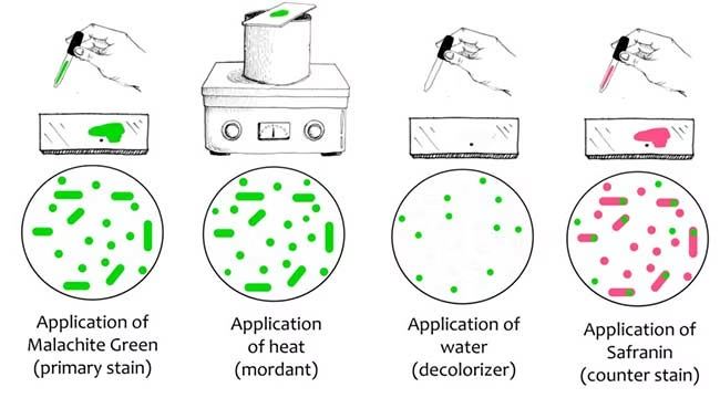
Stain with malachite green:
-
Place a few drops of malachite green solution on the smear.
-
Heat the slide gently by placing it over a steaming beaker of water or using a Bunsen burner. Steam the slide for about 5-10 minutes to facilitate the penetration of the malachite green into the spores. Make sure the dye does not evaporate.
-
-
Rinse with water:
-
After steaming, rinse the slide with distilled water to remove excess malachite green.
-
-
Counterstain with safranin:
-
Apply safranin to the slide and let it sit for 30 seconds to 1 minute. This will stain the vegetative cells a red or pink color.
-
-
Rinse and dry:
-
Rinse the slide with distilled water to remove excess safranin.
-
Blot the slide gently with a paper towel and allow it to air dry completely.
-
-
Examine under the microscope:
-
Use a compound light microscope with an oil immersion lens to observe the slide.
-
Spores will appear as green, oval or round structures inside or outside the red-stained vegetative cells.
-
Results
-
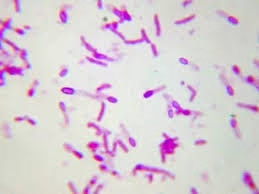
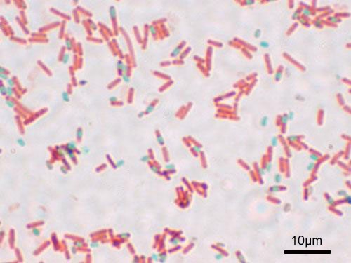
Spores: The spores will appear green and are often oval or round. They will either be inside the vegetative cells or free in the background. The green color comes from the malachite green dye, which penetrates the tough spore coat.
-
Vegetative Cells: The vegetative cells will appear red or pink, which is the result of the safranin counterstain. The contrast between the green spores and red cells makes it easy to identify the presence of spores.
Applications
-
Identifying Spore-Forming Bacteria: Spore staining is primarily used to identify spore-forming bacteria such as Bacillus and Clostridium species, which are important in both clinical diagnostics and industrial microbiology.
-
Diagnosis of Infections: Spore-forming bacteria like Clostridium tetani (causing tetanus) or Clostridium botulinum (causing botulism) can be diagnosed using this technique when suspected in clinical specimens.
-
Environmental Testing: In the food, water, and agricultural industries, spore staining helps detect potential contamination with spore-forming bacteria, which can be resistant to disinfectants and extreme environmental conditions.
-
Microbial Ecology Studies: Spore-forming bacteria are widespread in the environment, and spore staining is useful in environmental microbiology for studying these resilient organisms in soil, water, and other environmental samples.
-
Research: Spore staining is an important tool in microbiological research to study the life cycle, survival mechanisms, and environmental resistance of spore-forming bacteria.