Introduction
- Sporotrichosis is a fungal infection caused by Sporothrix schenckii, a dimorphic fungus that can infect humans and animals.
- This infection primarily affects the skin and subcutaneous tissues but can also involve the lymphatic system and other organs in more severe or disseminated cases.
- Sporotrichosis is often associated with occupations or activities that involve handling plant material, such as horticulturists, farmers, and gardeners, as the fungus is commonly found in soil, decaying vegetation, and plant material.
- The infection is usually contracted through traumatic inoculation of the fungus into the skin, often via cuts or abrasions from thorns, leaves, or other plant material.
- It is not typically transmitted from person to person.
Etiology
- The causative agent of sporotrichosis is Sporothrix schenckii, a dimorphic fungus, which means it can exist in two different forms depending on environmental conditions.
- In its environmental form, S. schenckii is a mold characterized by small conidia (spores) and long, branching conidiophores.
- When cultured at body temperature (37°C), S. schenckii converts to a yeast form, the pathogenic form in humans.
- This dimorphism is a key feature that allows S. schenckii to infect a host, as it can adapt to the temperature and conditions within the human body.
- schenckii is commonly found in soil, decaying plant matter, and on thorns of certain plants like roses, especially in areas with warm, humid climates.
- It can be isolated from animal tissues, particularly cats, which may act as reservoirs for the fungus, and transmission to humans may occur through contact with infected animals or their secretions.
Specimens
To diagnose sporotrichosis, various clinical specimens may be collected depending on the presentation of the disease:
- Skin and Subcutaneous Tissue: The most common site of infection is the skin, so samples are often obtained from skin lesions, ulcers, or abscesses. Skin scrapings or aspirates from pustules may be collected for analysis.
- Biopsy Samples: In chronic or deeper infections, such as when the infection has spread to lymph nodes or other tissues, biopsy samples of affected skin or tissues may be taken.
- Exudates or Pus: In cases of abscess formation, exudates or pus from the lesion may be collected to isolate the causative organism.
- Nail or Bone Specimens: In more severe or disseminated cases, samples from nails, bones, or other tissues may be required.
Direct Microscopic Examination
Direct microscopic examination plays a crucial role in diagnosing sporotrichosis. Common techniques include:
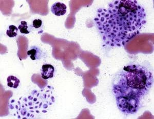
- KOH (Potassium Hydroxide) Preparation: Clinical specimens such as skin scrapings, pus, or biopsy material are treated with KOH, which dissolves keratin, making it easier to observe fungal elements under the microscope. The yeast form of S. schenckii is typically observed in infected tissue. These appear as round to oval cells with a narrow, constricted neck, resembling the typical “cigar-shaped” yeast cells.
- Histopathology: A biopsy specimen can be processed for histopathological examination. Special stains such as Periodic Acid-Schiff (PAS) or Gomori’s methenamine silver (GMS) stain can enhance the visibility of fungal elements in tissue sections. S. schenckii can be identified as yeast cells in tissue with associated granulomatous inflammation.
- Lactophenol Cotton Blue Staining: This stain visualizes fungal elements in culture. In the mold phase, S. schenckii produces small conidia and branching conidiophores that can be observed under the microscope.
- India Ink Staining: This technique can also be applied in some cases, although it is more commonly used for fungal infections like cryptococcosis. It can highlight the characteristic shape and morphology of S. schenckii in clinical specimens.
Culture and Identification
Culture is essential for confirming the diagnosis of sporotrichosis and identifying the specific fungal species involved. The following are key points for culturing Sporothrix schenckii:
- Culture Media: Sporothrix schenckii is cultured on Sabouraud dextrose agar (SDA) or Brain Heart Infusion (BHI) agar, both supporting fungal growth. Dermatophyte test media (DTM) can also be used. The fungus grows relatively slowly, with colonies typically appearing in 3-7 days.
- Incubation Conditions: Cultures are incubated at two different temperatures. At room temperature (25-30°C), the fungus grows in its mold form, producing a characteristic cottony or leathery colony with black, granular, or brownish pigmentation due to the production of conidia. The fungus converts to its yeast form at body temperature (37°C), producing creamy, moist colonies. The yeast form can also appear spherical to oval cells with a narrow neck, which are the “cigar-shaped” yeast cells.
- Colony Morphology: The morphology of the colony is a key characteristic. The colony appears white to light brown in the mold phase with a granular or powdery texture. The yeast form appears as white to cream-colored colonies that may be moist or smooth.
- Microscopic Examination: Microscopic examination of the fungal structures can provide important clues after colony growth. The yeast form (37°C) appears as cigar-shaped, narrow-necked cells. The mold form (at 25°C) produces conidia on branching conidiophores.
- Biochemical Identification: In some cases, biochemical tests or molecular methods, such as PCR or DNA sequencing, may be employed to identify S. schenckii and differentiate it from similar fungi. However, morphological characteristics are usually sufficient for identifying S. schenckii.
Other Laboratory Tests
Other laboratory tests that may be used in the diagnosis and management of sporotrichosis include:
- Serology: Although not routinely used in clinical practice, serological tests such as enzyme-linked immunosorbent assay (ELISA) can detect antibodies against S. schenckii in patients with chronic or disseminated infections. However, these tests are not highly sensitive and are typically more useful for epidemiological studies or cases where the diagnosis is uncertain.
- Polymerase Chain Reaction (PCR): PCR-based tests have been developed to detect Sporothrix schenckii DNA in clinical specimens, particularly in cases where culture and microscopy are inconclusive. PCR is more sensitive and specific than culture and can aid early diagnosis, especially in tissue samples.
- Immunohistochemistry (IHC): IHC may be used on biopsy samples to detect fungal antigens and confirm the presence of S. schenckii in tissues.
Pathogenesis
The pathogenesis of sporotrichosis involves the following steps:
- Inoculation: Infection begins when Sporothrix schenckii is introduced into the skin via trauma, such as a cut or abrasion, commonly through contact with contaminated plant material (e.g., thorns) or animal products. This is the most common route of infection.
- Transition to Pathogenic Form: Upon inoculation into the host, S. schenckii transforms from mold to yeast at body temperature. The yeast cells then proliferate in the tissues and induce an inflammatory response.
- Inflammatory Response: The infection typically starts as a localized, self-limited skin ulcer with a surrounding inflammatory response. Over time, it may progress to form multiple lesions along the lymphatic channels, creating a characteristic “sporotrichoid” pattern (linear, ascending skin lesions).
- Dissemination: In immunocompromised individuals or untreated cases, S. schenckii can disseminate beyond the skin to other organs such as the bones, joints, lungs, and central nervous system (CNS). This can lead to more severe forms of the disease, such as disseminated sporotrichosis.
- Chronic Granulomatous Inflammation: The immune system typically responds by forming granulomas around the fungal cells, leading to chronic inflammation. This may result in scarring, fibrosis, and disfigurement, particularly in prolonged infections.
Treatment of Sporotrichosis
The treatment of sporotrichosis depends on the severity and extent of the infection:
- Localized Cutaneous Sporotrichosis: For limited, localized skin infections, oral antifungal medications such as itraconazole or terbinafine are usually effective. These drugs inhibit ergosterol synthesis, disrupting the fungal cell membrane and leading to cell death. Treatment is generally continued for 3-6 months.
- Lymphocutaneous Sporotrichosis: In cases where the infection has spread along lymphatic channels, oral antifungals like itraconazole are used longer. In some cases, a combination of oral antifungals with local therapies, such as surgical drainage of abscesses, may be required.
- Disseminated Sporotrichosis: In severe, disseminated cases (especially in immunocompromised patients), a combination of itraconazole and amphotericin B may be necessary. Amphotericin B is a potent antifungal agent that can be administered intravenously for more severe infections, especially in patients with life-threatening diseases.
- Alternative Treatments: For patients intolerant to itraconazole or other oral antifungals, potassium iodide (KI) has been used, particularly in chronic or superficial sporotrichosis cases. Surgical intervention may be needed for draining abscesses or excising deeply involved tissue.
- Prevention: Preventive measures focus on avoiding direct contact with contaminated materials (such as plant thorns and soil). Wearing gloves and protective clothing can reduce the risk of exposure for workers in high-risk occupations.