
Introduction
- Trematodes, commonly known as flukes, are a group of parasitic flatworms belonging to the class Trematoda.
- These parasites are known for their leaf-shaped bodies and have complex life cycles involving one or more intermediate hosts.
- Trematodes infect various organs in the human body, such as the liver, intestines, lungs, and blood vessels.
Trematodes are divided into several groups based on the organs they infect, and they are often categorized as:
- Intestinal Flukes
- Blood Flukes (such as Schistosoma)
- Lung Flukes
Each group of trematodes has unique characteristics, life cycle, and clinical manifestations. Some of the most common fluke species that infect humans include:
- Intestinal Flukes (e.g., Fasciolopsis buski and Heterophyes heterophyes) affect the gastrointestinal tract.
- Blood Flukes (e.g., Schistosoma species), which affect the blood vessels, especially in the mesenteric veins or bladder veins.
- Lung Flukes (e.g., Paragonimus westermani), which affect the lungs.
Intestinal Flukes
Morphology of Intestinal Flukes
-
Fasciolopsis buski (Giant Intestinal Fluke):
- Shape: Leaf-shaped, flat, and broad with a smooth surface. The body is characterized by a wide, oval shape that helps it adhere to the intestinal lining.
- Size: Fasciolopsis buski is among the largest intestinal flukes, reaching lengths of 20–75 mm and widths of 20 mm.
- Suckers: It has two suckers—an oral sucker at the anterior (front) end, which is used to attach to the host’s intestinal wall, and a ventral sucker located near the mid-body. Both suckers help the fluke adhere to the tissues and acquire nutrients.
- Eggs: The eggs are oval, operculated (having a lid), and about 130-150 micrometers long. The operculum is characteristic and allows water to enter the egg for larval hatching.
-
Heterophyes heterophyes (Small Intestinal Fluke):
- Size: 1–2 mm long, much smaller than Fasciolopsis buski.
- Shape: Leaf-like and flattened, similar to Fasciolopsis, but smaller overall.
- Suckers: It has both an oral and ventral sucker for attachment to the host’s intestinal wall.
- Eggs: Eggs are small, around 20–30 micrometers long, oval, and operculated.
Life Cycle of Intestinal Flukes
- Eggs: The parasite’s eggs are passed in the feces of an infected person. When the eggs are released into the water, they hatch into miracidia.
- Miracidia: The miracidia (larvae) swim and must find a freshwater snail (the first intermediate host) to infect. The miracidium penetrates the snail’s tissues.
- Cercariae: Inside the snail, the miracidium develops into cercariae (another larval form). The cercariae leave the snail and swim freely in the water.
- Metacercariae: The cercariae attach to water plants, such as water chestnuts, and encyst to form metacercariae.
- Human Infection: Humans or animals ingest the metacercariae when eating contaminated, undercooked water plants. After ingestion, the metacercariae excyst in the intestine.
- Adult Flukes: The fluke larvae move from the intestines to the large intestine, where they mature into adult flukes. They attach to the intestinal wall, feed, and grow.
Diagnosis of Intestinal Flukes
- Stool Microscopy:
- The main diagnostic method is to look for the characteristic eggs in the feces of the infected person. The eggs have a distinctive operculum (a lid-like structure).
- The eggs may appear in stool during an active infection.
- Serology:
- Serological tests, such as ELISA (Enzyme-Linked Immunosorbent Assay), can detect antibodies produced in response to the parasite.
- Immunofluorescence or PCR:
- Sometimes, PCR (Polymerase Chain Reaction) or immunofluorescence assays may confirm infection by detecting parasite DNA or antigens in blood or stool samples.
Blood Flukes (Schistosoma)
Morphology of Schistosoma
-
Schistosoma mansoni (Mansoni Schistosomiasis):
- Male: Approximately 1.0–1.5 cm long, with a broad, cylindrical body. The male’s body is flattened on one side to accommodate the gynecophoral canal, a groove in which the female resides.
- Female: Long and slender compared to the male, measuring 1.3–2.0 cm long. She resides inside the male’s ventral groove.
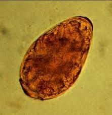
- Eggs: The eggs are oval with a lateral spine. They measure approximately 110–170 micrometers long. The lateral spine is a key feature for diagnosis.
-
Schistosoma haematobium (Haematobium Schistosomiasis):
- Male: Similar to S. mansoni, the male is about 1.0–1.5 cm in length, but its body is more rounded and not as flat.
- Female: Slender and smaller, and she resides in the gynecophoral canal of the male.
- Eggs: The eggs are oval with a terminal spine at the egg’s apex. They are 100–150 micrometers in size.
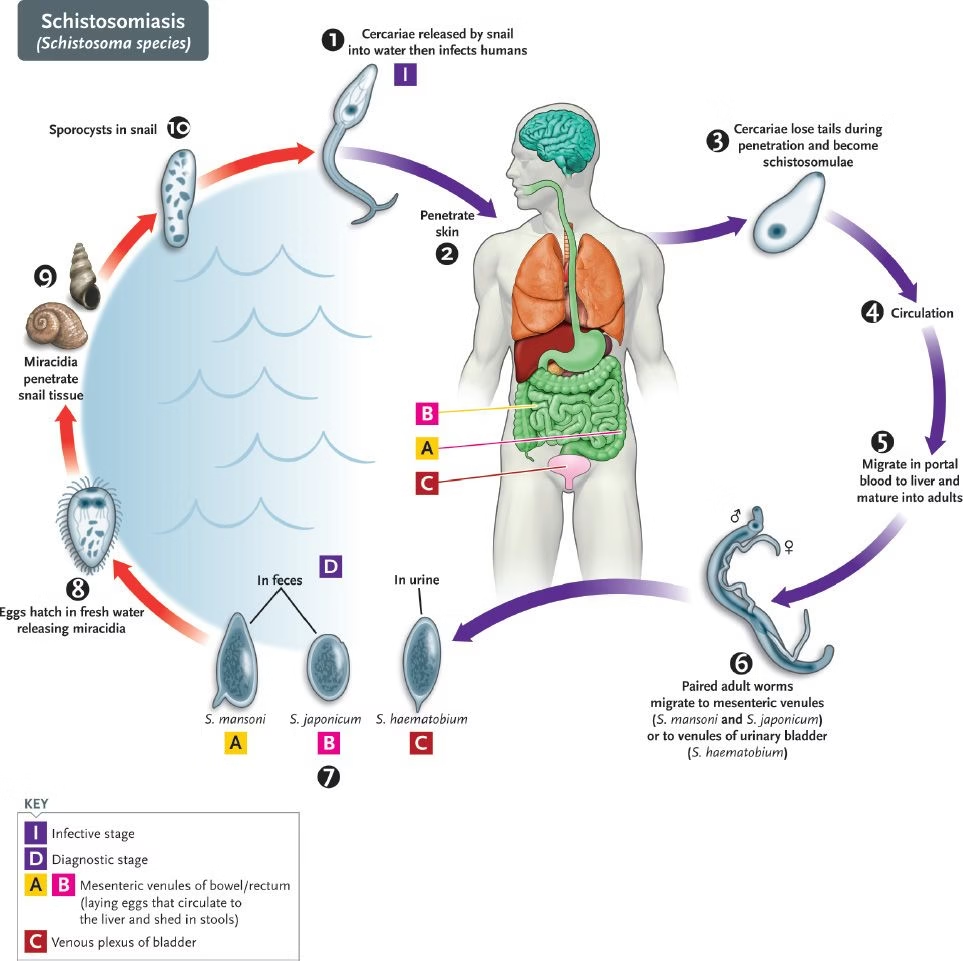
Life Cycle of Schistosoma
- Eggs: Eggs are passed in the feces or urine of infected humans.
- Miracidia: Eggs hatch in water, releasing miracidia, which infect freshwater snails.
- Cercariae: Inside the snail, the miracidium develops into cercariae. The snail releases these free-swimming cercariae into the water.
- Human Infection: The cercariae penetrate human skin when a person comes in contact with contaminated water.
- Schistosomulae: After penetration, the cercariae transform into schistosomulae, which travel via the bloodstream to the liver.
- Adult Flukes: The schistosomulae mature into adult male and female schistosomes in the liver. The adult flukes migrate to specific veins, like the mesenteric veins (S. mansoni) or vesical veins (S. haematobium), where they lay eggs.
- Eggs: The eggs exit the host’s body in feces (S. mansoni) or urine (S. haematobium), completing the life cycle.
Diagnosis of Schistosoma
-
Egg Identification in Stool or Urine:
- The most common diagnostic method is microscopic stool examination (for S. mansoni, S. japonicum) or urine (for S. haematobium) to find eggs with their characteristic spines.
- The lateral spine of S. mansoni and the terminal spine of S. haematobium are key identifiers.
-
Serological Tests:
- ELISA (Enzyme-Linked Immunosorbent Assay) can detect antibodies or antigens specific to Schistosoma.
- Indirect hemagglutination (IHA) tests may be used for detection in low-resource settings.
-
PCR:
- PCR can be used for molecular identification of Schistosoma DNA in blood, urine, or stool samples.
-
Imaging:
- In severe cases, ultrasound or CT scans may show organ damage, especially liver or spleen enlargement (hepatosplenomegaly) in chronic schistosomiasis.
Lung Flukes
Morphology of Paragonimus westermani (Lung Fluke)
- Shape: Oval, flattened, and leaf-like.
- Size: The adult fluke is about 7–12 mm long and 5–7 mm wide.
- Suckers: Oral sucker at the anterior end and a ventral sucker at the posterior end for attachment to lung tissue.
- Eggs: The eggs are oval, about 80–120 micrometers in size, and operculated (with a lid). These eggs are often found in sputum or stool.
Life Cycle of Paragonimus westermani
- Eggs: Eggs are passed out through infected individuals’ sputum (or stool). If swallowed, they are passed in feces.
- Miracidia: The eggs hatch into miracidia in water, which infect a freshwater snail (the intermediate host).
- Cercariae: After the snail develops, the cercariae are released into the water.
- Crustaceans: Cercariae infect freshwater crustaceans (such as crabs or crayfish), where they become metacercariae.
- Human Infection: Humans are infected when ingest raw or undercooked crustaceans containing the metacercariae.
- Migration to Lungs: After ingestion, the metacercariae exodes in the intestines and travels via the bloodstream to the lungs.
- Adult Flukes: The adults mature in the lung tissue, causing damage and inflammation. The adult flukes produce eggs, which are either coughed up in sputum or passed in stool.
Diagnosis of Paragonimus westermani
-
Microscopic Examination of Sputum:
- Eggs are often found in sputum (produced from coughing) or stool. The eggs have a characteristic operculum.
-
Chest Imaging:
- X-rays or CT scans may show lung lesions, including cavitary lesions typical of paragonimiasis.
-
Serological Tests:
- ELISA or Western blot can detect antibodies or antigens specific to Paragonimus.
-
PCR:
- PCR techniques can help detect Paragonimus DNA from sputum, stool, or serum samples.
Treatment for Trematode Infections
- Intestinal Flukes (e.g., Fasciolopsis buski):
- Praziquantel and triclabendazole are used to treat infections. These drugs help to kill adult flukes.
- Blood Flukes (Schistosoma):
- Praziquantel is the treatment of choice for all species of Schistosoma.
- Lung Flukes (Paragonimus westermani):
- Praziquantel is the treatment of choice for paragonimiasis.