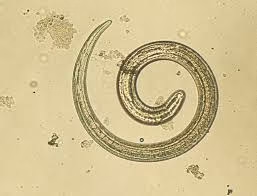
Habitat
- Definitive Habitat:
- The adult worms of Trichinella spiralis reside in the mucosa of the small intestine of their hosts, typically carnivorous or omnivorous animals (e.g., pigs, bears, and humans).
- Intermediate Habitat:
- The larvae form cysts in the striated muscles of the host, with a preference for active muscles such as the diaphragm, intercostals, tongue, extraocular muscles, and calf muscles.
- Encysted larvae can survive for years in host muscle, even in a calcified cyst.
Epidemiology
A. Geographic Distribution
- Trichinella spiralis is found worldwide. However, the prevalence depends on local dietary habits, food preparation practices, and animal husbandry.
- Common in regions with high pork consumption, particularly in China, Southeast Asia, Eastern Europe, and South America.
B. Hosts and Transmission
- Hosts:
- Definitive and intermediate hosts include humans, domestic animals (pigs), and wildlife (bears, foxes, wild boars, rodents).
- Mode of Transmission:
- Humans become infected by eating raw or undercooked meat (commonly pork or wild game) containing encysted larvae of Trichinella.
- Animals become infected through scavenging or feeding on infected meat or carcasses.
C. Outbreaks
- Associated with the consumption of improperly cooked pork, sausages, or wild game.
- Example: Outbreaks in hunters consuming wild boar or bear meat.
Morphology
A. Adult Worms
- Males:
- Size: ~1.4–1.6 mm in length and ~40–60 µm in diameter.
- Distinctive Feature: Lacks spicules but has two prominent caudal appendages used for copulation.
- Females:
- Size: ~3–4 mm long and ~60–80 µm in diameter.
- Viviparous (gives birth to live larvae).
B. Newborn Larvae (NBL)
- Size: ~80–120 µm in length and ~5–7 µm in diameter.
- Motile and capable of invading host tissues via the bloodstream.
C. Encysted Larvae
- Size: ~0.8–1 mm in length.
- Found encased within a capsule formed by host muscle tissue.
- Surrounded by a collagen-rich cyst wall, which calcifies over time.
Life Cycle
The life cycle of Trichinella spiralis is unique because a single host serves as both the definitive and intermediate host.
- Ingestion:
- Humans or animals consume raw or undercooked meat containing encysted larvae.
- Excystation:
- Gastric acid and pepsin in the stomach dissolve the cyst wall, releasing larvae into the stomach.
- Maturation in the Small Intestine:
- Larvae migrate to the intestinal mucosa and mature into adult worms within 24–48 hours.
- Male and female worms mate, and the females begin producing live newborn larvae.
- Larval Migration:
- Newborn larvae enter the lymphatic and circulatory systems, spreading to striated muscles.
- Larvae invade muscle fibers, where they grow and become encapsulated.
- Encystation in Muscle:
- Within 4–5 weeks, larvae encyst in striated muscle cells, forming nurse cell-larva complexes. These cysts become infective to new hosts.
- Cycle Continues:
- A new host must consume The infected muscle tissue for the life cycle to continue.
Important Notes:
- The lifecycle is self-limiting within humans since humans are typically dead-end hosts (i.e., humans are not eaten by other animals).
Pathogenicity
Trichinella spiralis causes trichinellosis (trichinosis), which progresses through three distinct phases:
A. Intestinal Phase (1–2 weeks post-infection)
- Caused by adult worms in the intestinal mucosa.
- Symptoms:
- Nausea, vomiting, diarrhea, abdominal pain, and mild fever.
- Occasionally asymptomatic, depending on the parasite load.
B. Systemic Phase (2–8 weeks post-infection)
- Caused by larval migration and encystation in muscle tissues.
- Symptoms:
- Fever, chills, headache.
- Muscle pain (myalgia), tenderness, and weakness, especially in heavily used muscles (e.g., diaphragm, extraocular muscles).
- Periorbital edema: Swelling around the eyes.
- Conjunctivitis and subconjunctival hemorrhages.
- Rash (urticarial or petechial).
C. Chronic Phase (after 8 weeks)
- Caused by encysted larvae in muscle tissues.
- Symptoms:
- Persistent muscle pain and fatigue.
- Severe cases may involve complications:
- Myocarditis: Inflammation of the heart muscles.
- Pneumonitis: Lung inflammation due to larval migration.
- Encephalitis or meningitis: If larvae migrate to the central nervous system (rare).
Complications:
- Severe infections with high larval loads may lead to multi-organ failure and, rarely, death.
Laboratory Diagnosis
A. Clinical History
- Recent consumption of raw or undercooked meat (e.g., pork or wild game).
- Symptoms such as fever, myalgia, and periorbital edema.
B. Laboratory Tests
- Serology:
- ELISA (Enzyme-Linked Immunosorbent Assay): Detects antibodies (IgG or IgM) specific to Trichinella.
- Western Blot: Confirms ELISA results with higher specificity.
- Muscle Biopsy:
- Histopathological examination of affected muscle reveals encysted larvae in cross-sections.
- Rarely performed today due to the advent of non-invasive serological methods.
- Complete Blood Count (CBC):
- Eosinophilia: A hallmark feature of trichinellosis.
- Elevated eosinophil count correlates with the severity of infection.
- Molecular Methods:
- PCR (Polymerase Chain Reaction): Detects Trichinella DNA in clinical specimens, providing a highly specific diagnosis.
- Imaging Studies:
- MRI or ultrasound may detect localized inflammation or calcified cysts in muscles.
Prevention and Control
A. Meat Preparation
- Cook meat to an internal temperature of at least 71°C (160°F).
- Freeze pork at -15°C for at least three weeks to kill larvae (not effective for some wild game species).
B. Public Health Measures
- Inspect slaughtered meat for encysted larvae.
- Educate people in endemic areas about the risks of consuming raw or undercooked meat.
Applications in Research
- Trichinella is studied extensively for its unique nurse cell-larva relationship in muscle fibers.
- Models of immune response to parasitic infections are often based on Trichinella spiralis.