
Introduction
- Periodic Acid-Schiff (PAS) stain is a widely used histochemical technique highlighting glycogen, mucopolysaccharides, glycoproteins, and other carbohydrate-rich tissue structures.
- It is commonly employed in pathology for diagnosing fungal infections, glycogen storage diseases, basement membrane abnormalities, and certain types of tumors.
- The PAS stain is particularly useful in identifying carbohydrate-laden components of cells and extracellular matrices, aiding in disease identification and classification.
Principle
- PAS staining is based on the oxidation of carbohydrates by periodic acid, which generates aldehyde groups.
- These aldehydes react with Schiff’s reagent to produce a magenta-colored complex, allowing visualization of carbohydrate-containing structures.
- The reaction is highly specific to polysaccharides, mucopolysaccharides, glycoproteins, and glycolipids, making PAS a valuable histopathological tool.
- The intensity of PAS staining depends on the concentration and distribution of carbohydrates within the tissue.
Requirements
- Microscope slides with properly fixed and sectioned tissue samples (paraffin-embedded or frozen sections)
- Staining rack for slide handling
- Glass beakers and cylinders for reagent preparation and application
- Distilled water for rinsing steps
- Timer to ensure precise incubation times
- Coverslips and mounting medium for slide preservation
- Light microscope for examination
Reagents
- Periodic Acid Solution (0.5-1%) – Oxidizes carbohydrates to aldehydes, making them reactive to Schiff’s reagent.
- Schiff’s Reagent – A colorless solution that reacts with aldehydes to form a magenta-colored complex upon binding.
- Sulfurous Acid or Sodium Metabisulfite Solution – Used to remove excess Schiff’s reagent, preventing non-specific staining.
- Hematoxylin (optional counterstain) – Stains nuclei blue, providing contrast for better visualization.
- Ethanol and Xylene – Used for dehydration, clearing, and slide preparation for microscopic evaluation.
- Diastase Enzyme (for PAS-D) – Used in diastase-PAS staining to digest glycogen, differentiating glycogen from other PAS-positive substances.
Procedure
- Deparaffinization and Hydration:
- Place tissue sections in xylene for 5-10 minutes to remove paraffin.
- Rehydrate slides through graded ethanol solutions (100%, 95%, 70%) down to distilled water.
- Oxidation:
- Immerse slides in 0.5-1% periodic acid solution for 5-10 minutes.
- Rinse well in distilled water to remove excess periodic acid.
- Schiff’s Reagent Reaction:
- At room temperature, Incubate slides in Schiff’s reagent for 10-15 minutes.
- Observe the tissue sections turning a faint pink color.
- Rinse slides in sulfurous acid or sodium metabisulfite solution (3 changes, 1-2 minutes each) to remove excess stain.
- Wash slides in running tap water for at least 5 minutes until the pink color intensifies.
- Counterstaining (if necessary):
- Stain slides with hematoxylin for 1-2 minutes to highlight nuclei.
- Rinse with tap water and blue in an alkaline solution if required.
- Dehydration and Mounting:
- Dehydrate slides through graded ethanol solutions (70%, 95%, 100%).
- Clear in xylene and mount with a permanent mounting medium.
- Microscopic Examination:
- Examine under a light microscope at appropriate magnifications.
Results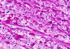
- PAS-positive structures: Magenta/purple (glycogen, mucins, basement membranes, fungal cell walls, some types of tumor cells)
- PAS-negative structures: Unstained or background counterstained with hematoxylin (blue nuclei)
- PAS-D (Diastase PAS) results:
- Glycogen-positive areas disappear after diastase treatment (confirming glycogen content).
- Other PAS-positive components remain unchanged (e.g., mucins, fungal walls, basement membranes).
Applications
- Identification of glycogen storage diseases (PAS-positive, diastase-sensitive glycogen deposits).
- Diagnosis of fungal infections (fungal cell walls stain magenta with PAS).
- Detection of mucin-secreting adenocarcinomas.
- Examination of renal and hepatic diseases involving basement membrane changes.
- Analysis of muscle and connective tissue disorders involving glycoproteins.
Advantages
- Highly sensitive for detecting glycogen, mucins, and polysaccharides.
- Useful in diagnosing fungal infections, basement membrane abnormalities, and certain tumors.
- Provides good contrast when combined with hematoxylin counterstaining.
- It can be used alongside diastase digestion (PAS-D) to confirm glycogen presence.
Disadvantages
- Non-specific staining can occur if not properly controlled.
- Cannot distinguish between different types of carbohydrates without additional techniques.
- Schiff’s reagent is light-sensitive and must be stored carefully to maintain its efficacy.
- Requires well-fixed tissue samples, as improper fixation can affect staining quality.
Limitations
- Does not differentiate between various types of polysaccharides and glycoproteins.
- It may produce false positives due to contaminants or improper fixation.
- Additional confirmatory tests (e.g., diastase digestion) are required to identify glycogen specifically.
- Staining results may vary based on fixation and tissue processing techniques.