
General features of Cartilage
- Histology of Cartilage is an avascular connective tissue that forms the “skeletal” basis of some parts of the body, e.g. the auricle of the ear, or the lower part of the nose.
- Cartilage develops from mesenchyme.
- They are not rigid but firm enough to maintain the form of the structure.
- Cartilage contains cells and fibres embedded in a homogeneous ground substance.
- Cells are relatively less and are called as chondrocytes.
Composition of Cartilage
Cartilage = Chondrocytes + Extracellular matrix (ground substance + fibers)
Like ordinary connective tissue, cartilage is made up of:
Cells—chondrocytes
Intercellular/extracellular matrix
Fibers—collagen/elastic/reticular fibers.
- Mature cells of cartilage are called as chondrocytes.
- They vary in size and shape with the degree of maturation.
- Mature cells are large and rounded.
- They lie in spaces (or lacunae) present in the matrix.
Ground Substance
- Ground substance forms major component and makes up to 95% of volume of the cartilage.
- The ground substance is made up of complex molecules containing proteins and carbohydrates (proteoglycans).
- The carbohydrates are chemically glycosaminoglycans (GAGs).
Fibers of Cartilage
- The collagen fibers present in cartilage are (as a rule) chemically distinct from those in most other tissues.
- They are described as type II collagen.
- However, type I collagen is present in fibrocartilage and perichondrium.
- Elastic cartilage contains elastic fibers.
Type of Cartilage
Hyaline Cartilage
- Hyaline cartilage is so called because it is translucent (hyalos = glass).
- Its matrix appears to be homogeneous, but using special techniques, many collagen fibers can be demonstrated.
- Hyaline cartilage has been compared to a tyre.
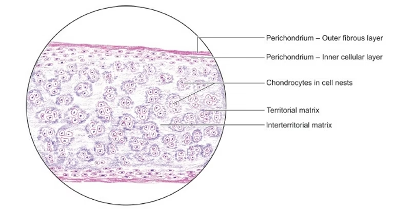
- In hematoxylin and eosin-stained preparations, the matrix is stained blue, i.e. it is basophilic.
- The matrix around the cells is darkly stained, called as capsular matrix and around the isogenous cell group as the territorial/lacunar matrix.
Functions
- By forming a structural framework that provides support to various structures
- Form a cartilage model for developing the fetal skeleton
- Forms articular cartilage.
Elastic Cartilage
- Elastic cartilage (or yellow fibrocartilage) is similar in many ways to hyaline cartilage.
- The main difference between hyaline cartilage and elastic cartilage is that instead of collagen fibers, the matrix contains numerous branching and anastomosing elastic fibers that form a network.
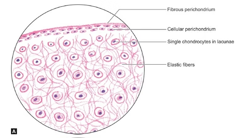
- The fibers are difficult to see in hematoxylin and eosin-stained sections, but they can be clearly visualized if special methods for staining elastic fibers are used.
- The surface of elastic cartilage is covered by perichondrium.
- In contrast to hyaline cartilage, elastic cartilage does not undergo calcification.
Function
- Provides elasticity (flexibility) to the structures.
Fibrocartilage
- Fibrous cartilage (also called white fibrocartilage) looks very much like dense fibrous tissue.
- Since fibrocartilage is a transitional tissue between dense connective tissue and hyaline cartilage, lack of perichondrium helps in its proper blending with surrounding structure.
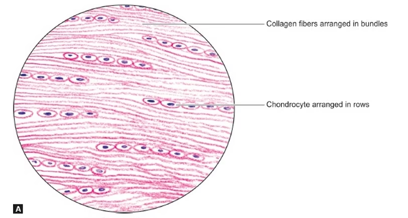
- The chondrocytes are similar to those of hyaline cartilage but are arranged in single rows or in isogenous groups.
Functions
- Offers resistance to both compression and shearing forces
- They act as shock absorbers.
Applied Aspects
-
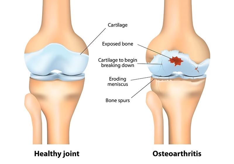
Joint Cartilage (Osteoarthritis): Damage to hyaline cartilage in joints can lead to conditions like osteoarthritis. Since cartilage is avascular, its repair is limited, making degenerative diseases challenging to treat.
-
Cartilage Transplants/Regeneration: Research into stem cell therapies and tissue engineering aims to regenerate or replace damaged cartilage, especially in joint diseases like arthritis.
-
Cartilage in the Respiratory System: Cartilage in the trachea and bronchi prevents airway collapse and ensures smooth airflow.
-
Developmental Biology: Cartilage serves as a precursor to bone in the fetal skeleton, and its study provides insights into skeletal development and disorders.
-
Injury Repair and Regeneration: In some cases, cartilage injuries, such as those seen in athletes, can be repaired using regenerative techniques like microfracture surgery, autologous chondrocyte implantation, or stem cell therapy.