
Introduction
-
Pathology is one of the most important and core branches of medical science.
-
It is the study of disease, including:
-
How diseases develop
-
How they affect the body
-
How they can be diagnosed
-
-
Pathology helps in diagnosis through:
-
Laboratory testing
-
Examination of tissues, cells, blood, and body fluids
-
-
Pathology acts as a bridge between:
-
Basic medical sciences (Anatomy, Physiology, Biochemistry)
-
Clinical medicine
-
-
In modern healthcare, almost every diagnosis is confirmed:
-
Directly or indirectly
-
With the help of pathology investigations and reports
-
History of Pathology
The history of pathology is long and fascinating. It has evolved from simple observation of the body to highly advanced molecular and genetic diagnostic techniques.
1 Ancient Period
-
In ancient times, diseases were believed to be caused by evil spirits, curses, or supernatural forces.
-
Egyptian and Greek civilizations started early medical documentation.
-
Hippocrates (460–370 BC) is considered the Father of Medicine.
-
He suggested that diseases occur due to imbalance in body fluids (humoral theory: blood, phlegm, black bile, yellow bile).
-
2 Roman Period
-
Galen (129–216 AD) expanded the humoral theory.
-
He contributed to anatomy and physiology through animal dissections.
-
However, the understanding of disease remained limited.
3 Middle Ages
-
During this period, medical progress slowed due to restrictions on human dissection.
-
Diseases like plague spread rapidly.
-
Still, hospitals and medical learning began developing gradually.
4 Renaissance Period (14th–17th Century)
This period marked a major turning point.
-
Human dissections were allowed and encouraged.
-
Andreas Vesalius (1514–1564) corrected many anatomical mistakes.
-
This helped in correlating disease with structural changes in the body.
5 Development of Autopsy-Based Pathology
-
Giovanni Morgagni (1682–1771) is known as the Father of Anatomical Pathology.
-
He correlated clinical symptoms with post-mortem (autopsy) findings.
-
This created the foundation of modern pathology.
6 Cellular Pathology (19th Century)
-
Rudolf Virchow (1821–1902) is called the Father of Modern Pathology.
-
He introduced the concept:
“Omnis cellula e cellula”
(All cells arise from pre-existing cells) -
He proved that disease begins at the cellular level, not only in organs.
7 Rise of Microbiology and Infectious Disease Pathology
-
Louis Pasteur and Robert Koch proved that microorganisms cause diseases.
-
Koch’s postulates helped establish infectious disease pathology.
8 Modern Era (20th Century to Present)
Pathology has expanded massively with:
-
Histopathology
-
Clinical pathology
-
Immunohistochemistry
-
Molecular pathology
-
Genetic testing
-
Digital pathology and AI
Today, pathology is not only used for diagnosis but also for:
-
Prognosis
-
Treatment monitoring
-
Personalized medicine
Basic Definitions in Pathology
Understanding the basic terms is essential for every medical and paramedical student.
1 Pathology
Pathology is the scientific study of disease, including:
-
Cause (etiology)
-
Mechanism (pathogenesis)
-
Structural changes (morphology)
-
Functional changes and clinical effects
2 Disease
A disease is a condition in which normal body structure or function is disturbed.
3 Lesion
A lesion is any abnormal change in tissue or organ caused by disease.
4 Diagnosis
Diagnosis is the identification of a disease based on:
-
Symptoms
-
Signs
-
Laboratory findings
-
Histopathological examination
5 Etiology
Etiology means the cause of disease.
Example:
-
Bacteria → tuberculosis
-
Smoking → lung cancer
-
Vitamin deficiency → scurvy
6 Pathogenesis
Pathogenesis is the mechanism and sequence of events that lead to disease development.
7 Morphology
Morphology refers to the structural changes in tissues or cells due to disease.
8 Prognosis
Prognosis means the expected outcome of the disease.
Familiarisation
Pathology has its own set of frequently used terms. Knowing them improves clinical and lab understanding.
1 Inflammation
Inflammation is the body’s response to injury or infection.
Types:
-
Acute inflammation: sudden, short duration (e.g., abscess)
-
Chronic inflammation: long duration (e.g., tuberculosis)
2 Necrosis
Necrosis is irreversible cell death due to injury.
Common types:
-
Coagulative necrosis (myocardial infarction)
-
Liquefactive necrosis (brain infarct)
-
Caseous necrosis (TB)
3 Apoptosis
Apoptosis is programmed cell death (controlled and normal).
4 Degeneration
Degeneration means reversible cell injury where cells show abnormal changes but may recover.
Example:
-
Fatty change in liver
5 Hyperplasia
Increase in number of cells.
Example:
-
Benign prostatic hyperplasia
6 Hypertrophy
Increase in size of cells.
Example:
-
Left ventricular hypertrophy in hypertension
7 Metaplasia
Reversible change where one adult cell type is replaced by another.
Example:
-
Squamous metaplasia in smokers
8 Dysplasia
Disordered growth of cells, considered a premalignant condition.
Example:
-
Cervical dysplasia
9 Neoplasia
New abnormal growth of tissue (tumor).
Types:
-
Benign tumor
-
Malignant tumor (cancer)
10 Biopsy
Removal of tissue from living body for microscopic examination.
11 Autopsy
Examination of dead body to determine cause of death and study disease.
Branches of Pathology
Pathology is broadly divided into:
Anatomical Pathology
Deals with:
-
Tissues
-
Organs
-
Cells
Includes:
-
Histopathology
-
Cytopathology
-
Autopsy pathology
Clinical Pathology (Laboratory Medicine)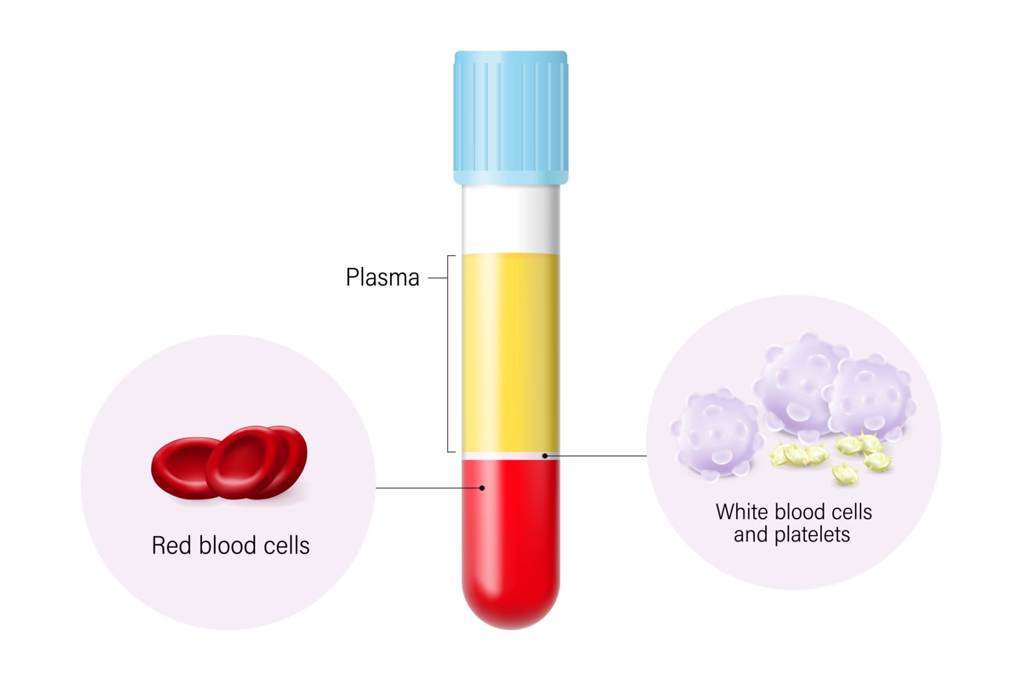
Deals with:
-
Blood
-
Urine
-
Body fluids
-
Biochemical tests
Includes:
-
Hematology
-
Clinical biochemistry
-
Microbiology
-
Immunology
Techniques Used in Pathology
Modern pathology depends on multiple techniques for accurate diagnosis.
Gross Examination
-
The first step in tissue examination.
-
The specimen is observed with naked eyes.
-
Features noted:
-
Size
-
Shape
-
Color
-
Consistency
-
Cut surface appearance
-
Example:
-
Tumor mass in breast biopsy
Fixation
Fixation preserves tissue and prevents decomposition.
Common fixative:
-
10% Neutral Buffered Formalin
Purpose:
-
Maintains tissue architecture
-
Prevents autolysis and putrefaction
Tissue Processing
After fixation, tissue is processed in:
-
Dehydration (using alcohol)
-
Clearing (xylene)
-
Paraffin wax embedding
This allows thin section cutting.
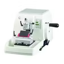
Microtomy
-
Cutting thin tissue sections (3–5 microns) using microtome.
-
Sections are placed on glass slides.
Staining
Staining highlights cells and tissues under microscope.
(A) Routine Stain
Hematoxylin and Eosin (H&E) stain
-
Hematoxylin stains nucleus (blue/purple)
-
Eosin stains cytoplasm (pink)
(B) Special Stains
Used when H&E is not enough.
Examples:
-
PAS stain → fungi, glycogen
-
Ziehl-Neelsen stain → acid-fast bacilli (TB)
-
Gram stain → bacteria
-
Congo red → amyloid
Cytology Techniques
Cytology studies individual cells.
Types:
-
Pap smear (cervical cancer screening)
-
FNAC (fine needle aspiration cytology)
FNAC is commonly used for:
-
Thyroid nodules
-
Breast lumps
-
Lymph nodes
Hematology Techniques
Used for blood-related diagnosis.
Includes:
-
Complete Blood Count (CBC)
-
Peripheral smear examination
-
ESR
-
Coagulation tests (PT, APTT)
Microbiological Techniques
Used to identify infectious agents.
Includes:
-
Culture and sensitivity testing
-
Microscopy
-
Serological tests
-
Molecular tests (PCR)

close up shot of microscope at the blood laboratory and red blood cells under microscope take with art lighting and blue filter
Immunohistochemistry (IHC)
IHC uses antibodies to detect specific proteins in tissues.
Applications:
-
Tumor typing
-
Identifying tumor origin
-
Prognostic markers
Example:
-
ER/PR in breast cancer
-
HER2 status
Molecular Pathology
Modern diagnostic technique that detects genetic changes.
Includes:
-
PCR
-
RT-PCR
-
DNA sequencing
-
FISH (Fluorescence in situ hybridization)
Applications:
-
Detecting mutations in cancers
-
Viral load testing
-
Genetic diseases
Electron Microscopy
Used for very high magnification.
Applications:
-
Kidney diseases (glomerular pathology)
-
Some viral infections
-
Muscle disorders
Flow Cytometry
A powerful technique for analyzing cell markers.
Used mainly for:
-
Leukemia and lymphoma diagnosis
-
Immunophenotyping
Importance of Pathology in Modern Healthcare
Pathology is essential because it helps in:
-
Early diagnosis of diseases
-
Confirmation of clinical diagnosis
-
Cancer detection and staging
-
Monitoring treatment response
-
Identifying infectious organisms
-
Guiding personalized therapy