Introduction
- Bacteria are unicellular prokaryotic microorganisms that show remarkable diversity in shape, size, and arrangement.
- Morphology is one of the fundamental criteria used in microbiology for classification, identification, and diagnosis.
- Understanding bacterial morphology helps students interpret Gram staining results, culture characteristics, and microscopic findings in clinical samples.
Bacterial morphology is studied under:
-
Shape (morphological type)
-
Size
-
Arrangement
-
Special structural modifications
Size of Bacteria
Bacteria are measured in micrometers (µm).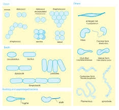
Average Size
-
Most bacteria range between 0.5–1.0 µm in width
-
Length varies from 1–5 µm
Examples
-
Cocci: 0.5–1 µm diameter
-
Bacilli: 1–4 µm length
-
Spirochetes: Up to 10–20 µm long
Smallest Bacteria
-
Mycoplasma: 0.2–0.3 µm
Largest Bacteria
-
Thiomargarita namibiensis: up to 750 µm (visible to naked eye)
Importance of Size
-
Influences nutrient absorption
-
Affects antibiotic penetration
-
Determines filtration ability
-
Important in microscopy resolution
Morphological Types of Bacteria
Bacteria are classified morphologically into the following major types:
A. Cocci (Spherical Bacteria)
-
Spherical or oval shape
-
Usually 0.5–1 µm in diameter
-
May occur singly or in characteristic arrangements
Arrangements of Cocci
-
Diplococci – in pairs
-
Streptococci – in chains
-
Staphylococci – in grape-like clusters
-
Tetrads – groups of four
-
Sarcinae – cubical packets of eight
Clinical Importance
Arrangement helps in identification during Gram staining.
B. Bacilli (Rod-Shaped Bacteria)
-
Cylindrical or rod-shaped
-
Length: 1–4 µm
-
Width: 0.5–1 µm
Variations
-
Straight bacilli
-
Coccobacilli (short rods)
-
Fusiform bacilli (spindle-shaped)
-
Filamentous bacilli
Arrangements
-
Single rods
-
Chains (streptobacilli)
-
Palisade arrangement (Chinese letter pattern)
C. Spiral Forms
Types
-
Vibrio
-
Comma-shaped
-
Slightly curved rod
-
-
Spirillum
-
Rigid spiral shape
-
Few helical turns
-
-
Spirochetes
-
Flexible spiral
-
Multiple coils
-
Motile
-
Size
-
Length varies from 5–20 µm
-
Thin and delicate
3. Pleomorphic Bacteria
Causes
-
Lack of cell wall
-
Nutritional deficiency
-
Aging culture
Example
-
Mycoplasma
-
Coryneform bacteria
Special Morphological Forms
A. Filamentous Bacteria
-
Long thread-like structures
-
Resemble fungal hyphae
-
Found in soil and some infections
B. Branching Bacteria
-
Show branching pattern
-
Seen in certain pathogenic species
C. Capsule Formation
-
Some bacteria produce outer gelatinous covering
-
Visible with special staining
D. Spore-Forming Bacteria
-
Produce endospores
-
Spores may be central, subterminal, or terminal
Arrangement of Bacteria
The arrangement of bacteria refers to the pattern in which bacterial cells are organized after cell division. While the shape (cocci, bacilli, spiral) defines morphology, the arrangement depends on: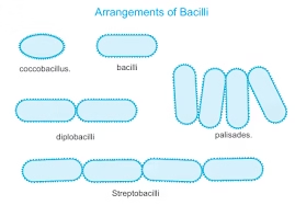
-
The plane of cell division
-
Whether daughter cells remain attached
-
The presence of capsule or extracellular substances
Arrangement is especially important in Gram staining interpretation and preliminary laboratory identification.
Arrangement of Cocci (Spherical Bacteria)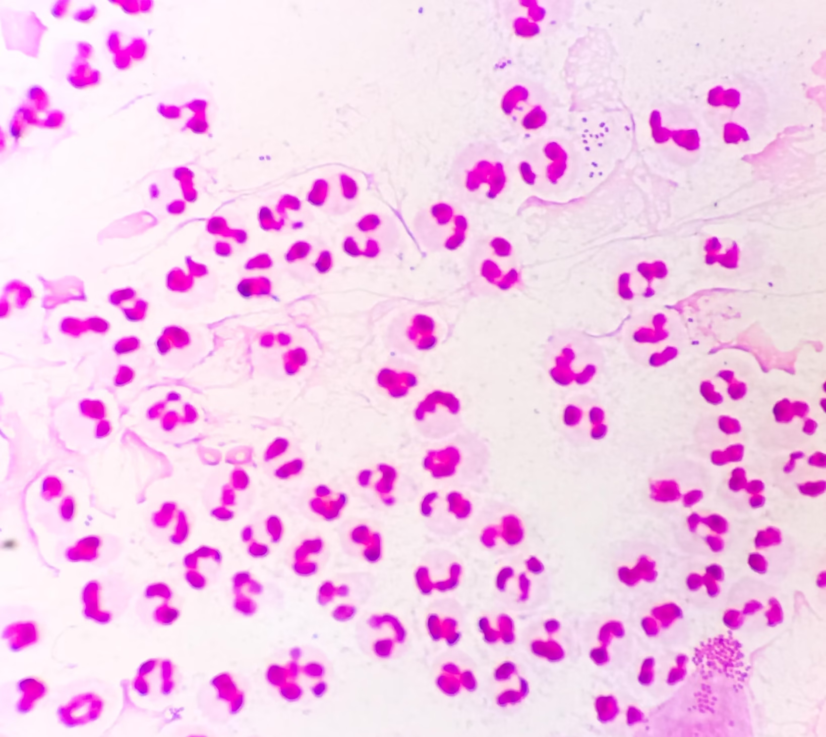
1. Diplococci
-
Division in one plane
-
Cells remain in pairs
-
Example appearance: Two cocci attached
2. Streptococci
-
Repeated division in one plane
-
Form chains
-
Cells appear like beads on a string
3. Staphylococci
-
Division in multiple planes
-
Irregular grape-like clusters
4. Tetrads
-
Division in two perpendicular planes
-
Groups of four cells
5. Sarcinae
-
Division in three perpendicular planes
-
Cubical packets of eight or multiples of eight
Arrangement of Bacilli (Rod-Shaped Bacteria)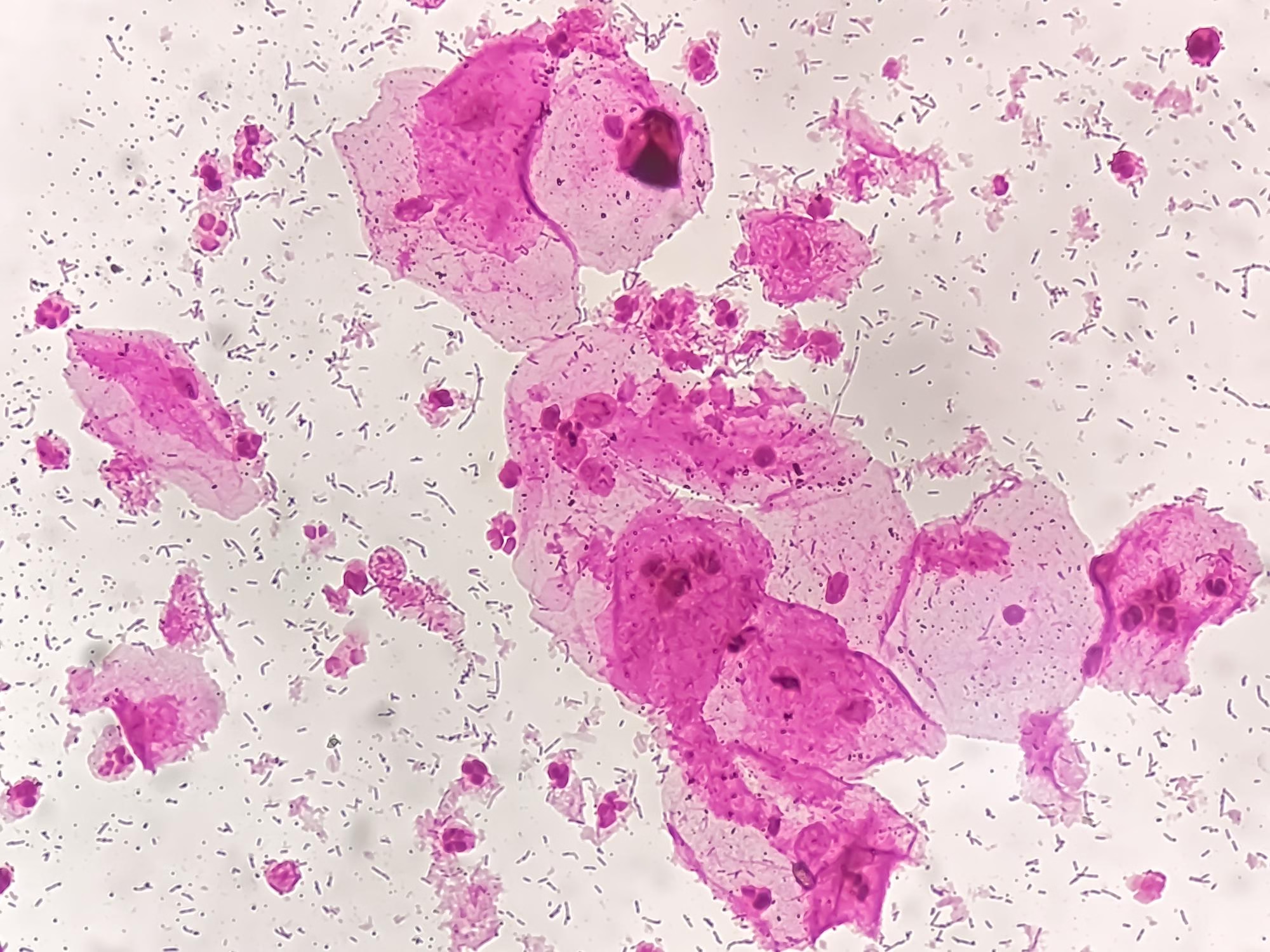
1. Single Bacilli
-
Occur individually
-
Most common arrangement
2. Diplobacilli
-
Occur in pairs
3. Streptobacilli
-
Form chains of rods
4. Palisade Arrangement
-
Side-by-side arrangement
-
Appears like “Chinese letters” or picket fence
5. Coccobacilli
-
Short, oval-shaped rods
-
Appear intermediate between cocci and bacilli
Arrangement of Spiral Bacteria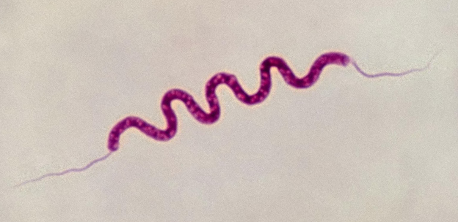
1. Vibrio
-
Comma-shaped
-
Usually single
2. Spirillum
-
Rigid spiral
-
Occur singly
3. Spirochetes
-
Flexible spiral
-
Occur singly
Factors Affecting Bacterial Morphology
1. Nutritional Factors
-
Availability of nutrients affects bacterial growth and structure.
-
Nutrient deficiency may cause:
-
Smaller cell size
-
Irregular shapes
-
Pleomorphism
-
-
Rich media generally produce normal morphology.
2. Age of Culture
-
Young cultures show typical morphology.
-
Old cultures may show:
-
Degenerative changes
-
Irregular staining
-
Cell shrinkage
-
Pleomorphic forms
-
This is why fresh cultures are recommended for Gram staining.
3. Temperature
-
Optimal temperature maintains normal morphology.
-
High temperature may cause:
-
Cell distortion
-
Abnormal elongation
-
-
Low temperature may slow growth and alter size.
Extreme temperatures damage cellular structure.
4. pH of the Environment
-
Most bacteria prefer neutral pH.
-
Acidic or alkaline conditions can cause:
-
Shape irregularities
-
Cell wall damage
-
Reduced size
-
5. Antibiotic Exposure
-
Antibiotics affecting cell wall synthesis may cause:
-
Swelling
-
Spheroplast formation
-
Filamentation
-
-
Sublethal doses may produce abnormal elongated cells.
Morphological changes can indicate antibiotic stress.
6. Osmotic Pressure
-
Hypertonic solutions cause cell shrinkage (plasmolysis).
-
Hypotonic solutions may cause swelling.
-
Changes in osmotic balance affect cell shape and integrity.
7. Oxygen Availability
-
Aerobic and anaerobic conditions influence metabolic activity.
-
Improper oxygen levels may affect growth rate and morphology.
8. Genetic Factors
-
Mutations may alter:
-
Cell shape
-
Cell wall structure
-
Arrangement pattern
-
-
Some bacteria are naturally pleomorphic due to genetic traits.
9. Mechanical and Physical Stress
-
Excessive shaking
-
Rough handling
-
Improper smear preparation
These may distort cells during microscopy.
10. Chemical Agents and Disinfectants
-
Exposure to chemicals can:
-
Damage cell wall
-
Cause cell lysis
-
Alter staining properties
-
Importance of Studying Bacterial Morphology
1. First Step in Laboratory Identification
-
Morphology is the initial observation after Gram staining.
-
Shape and arrangement immediately narrow down possible organisms.
-
Example: Gram-positive cocci in clusters suggest staphylococci.
This allows rapid presumptive diagnosis before culture confirmation.
2. Rapid Clinical Decision-Making
-
In life-threatening infections (e.g., meningitis, septicemia), immediate microscopic findings guide empirical antibiotic therapy.
-
Morphology provides quick differentiation between Gram-positive and Gram-negative bacteria.
3. Basis for Classification and Taxonomy
-
Early bacterial classification relied mainly on morphology.
-
Even today, morphology complements biochemical and molecular identification.
-
Shape and structural features remain taxonomic markers.
4. Understanding Pathogenic Mechanisms
-
Bacterial shape influences motility and tissue invasion.
-
Spiral bacteria move efficiently through mucus layers.
-
Capsule presence enhances virulence.
-
Spore formation ensures survival in adverse conditions.
Morphology is directly linked to pathogenic potential.
5. Guidance for Further Laboratory Testing
-
Observed morphology helps select appropriate culture media.
-
Guides biochemical test selection.
-
Indicates need for special staining techniques (e.g., spore stain, capsule stain).
6. Detection of Special Structural Features
Morphological study helps identify:
-
Spores (central, terminal, subterminal)
-
Capsules
-
Flagella
-
Pleomorphic variations
These features are important for diagnosis and treatment planning.
7. Monitoring Antibiotic Effect
-
Certain antibiotics cause morphological changes.
-
Abnormal shapes may indicate cell wall damage.
-
Helps assess bacterial response to treatment.
8. Epidemiological and Research Importance
-
Helps track bacterial strains in outbreaks.
-
Useful in environmental microbiology.
-
Important in teaching and basic microbiology research.