Introduction
- Blood vessels deliver nutrients, oxygen and hormones to cells of the body and remove metabolic waste products and CO2
from them through blood. - Exchange of these substances takes place at the capillary level.
Types of blood vessels
Histologically, there are five main types of blood vessels. They are:
1. Arteries
(a) Large artery (elastic artery)
(b) Medium-sized artery (muscular artery)
2. Arterioles
3. Capillaries
(a) Continuous capillary
(b) Fenestrated capillary
(c) Sinusoidal capillary
4. Venules
5. Veins
(a) Medium-sized vein
(b) Large vein
Structure
All blood vessels have the same basic structure. Each has three coats or tunics, namely,
(a) Tunica intima– It is composed of lining endothelium (simple squamous epithelium) and subendothelial connective tissue.
(b) Tunica media– This layer is made of smooth muscle and connective tissue.
(c) Tunica adventitia– It is constituted of fibroelastic connective tissue.
Arteries
Features
- Arteries are thick-walled blood vessels that carry blood from the heart to the capillaries.
- They divide repeatedly like a branch of a tree and gradually become smaller in size.
- However, their luminal surface is increased many times (800-fold) compared to that of a large artery (aorta).
- This causes a decrease in the rate of blood flow, facilitating the exchange of substances through the capillaries.
Structure
The arteries are subdivided into the following types based on their structure and size:
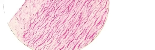
1. Large/Elastic/Conducting artery
Example, aorta and its branches:
- It conducts blood from the heart.
- Thickness of its wall is about one-tenth of the luminal diameter that varies.
- The presence of elastic fibres in the wall allows it to expand during contraction (systole) and to recoil during relaxation
(diastole) of the heart. This maintains necessary blood pressure, and thus permits the blood to fl ow more evenly through
the other arterial channels.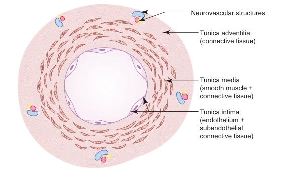
- The following are the layers of large arteries:
(a) Tunica intima (100 μm thick)
– It includes endothelium and subendothelial connective tissue.
– Subendothelial tissue contains fibrocytes, macrophages and smooth muscle-like cells called myointimal cells.
The fibres (collagen and elastic) in it are longitudinally oriented.
– Tunica intima is demarcated from tunica media by a poorly defi ned fenestrated internal elastic lamina.
(b) Tunica media
– It is mainly made of about 40–70 layers of fenestrated elastic laminae arranged circularly. Hence the name
elastic artery.
– Between elastic laminae, it contains smooth muscle cells and collagen fibres embedded in a basophilic matrix
rich in chondroitin sulphate.
– The outermost elastic lamina is thickened and called the external elastic lamina.
(c) Tunica adventitia
– It is composed of fibroelastic connective tissue carrying small blood vessels (vasa vasorum) and unmyelinated sympathetic fibres.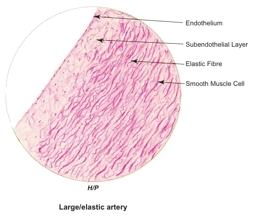
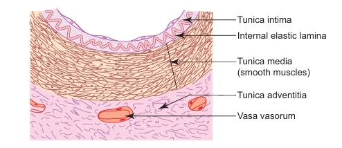
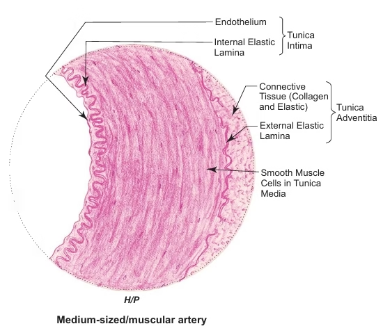
2. Medium-sized/Muscular/Distributing artery
Example: branches of the external carotid artery, radial and ulnar arteries:
- It distributes blood to various parts of the body.
- Its wall thickness is about one-fourth of the luminal diameter.
- The presence of smooth muscle in its wall helps to control the flow and pressure of blood through vasoconstriction or vasodilatation.
- The three layers of the wall (Fig. 10.2) are as follows:
(a) Tunica intima
– It is made of endothelium and internal elastic lamina (no subendothelium).
– The internal elastic lamina is a bright refractile membrane thrown into wavy folds due to contraction of
smooth muscle in the media.
(b) Tunica media
– It consists mainly of smooth muscle cells arranged circularly (about 40 layers). Hence the name muscular artery.
– It also contains elastic and a few collagen fibres intermixed with smooth muscle cells.
(c) Tunica adventitia
– The inner part of the tunica adventitia contains more elastic than collagen fibres, and it includes the external
elastic lamina.
– The middle part contains collagen and elastic fibres running longitudinally.
– The outer part is made of loose connective tissue that merges with the surrounding areolar tissue and
contains vasa vasorum and unmyelinated sympathetic nerve fibres.
Cappilaries
Feature
- Arterioles break up into small blood vessels called capillaries.
- Capillaries are often referred to as exchange vessels, because they are involved in the exchange of gases, nutrients and metabolites between blood and tissue.
- Tissues with high metabolic rates have an abundant capillary network (e.g. kidney, liver, cardiac muscle).
Structure
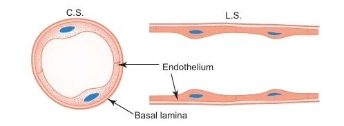
- The lumen of a typical capillary is about 7–9 μm wide (equal to the diameter of an erythrocyte) and is lined by endothelial cells, which are two or three in number on cross section of vessel and form its tunica intima.
- The margin of endothelial cells are held together by tight and gap junctions. Numerous pinocytotic vesicles are seen in the cytoplasm.
- They are involved in transporting material across the endothelial lining in either direction.
- Pericytes or adventitial cells are occasionally seen within the basement membrane of the endothelium constituting the tunica media.
- These cells contain contractile fi laments in the cytoplasm and can transform into other cells.
- A thin layer of collagen fi bres that surround the capillaries form the tunica adventitia.
- Capillaries are divided into following three types depending on the nature of the endothelium:
1. Continuous or somatic capillary
-
- It is the commonest type of capillary present in connective tissue, muscle, brain, lung, etc.
- The endothelial cells form a continuous lining of the capillary.
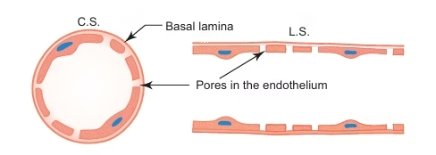
2. Fenestrated or visceral capillary
-
- This is characterised by the presence of tiny pores in the endothelial cells.
- These pores are often closed by a thin diaphragm (thinner than the cell membrane) and allow dissolved substances and macromolecules to pass through slowly.
- The permeability of fenestrated capillary is much greater than that of continuous capillary.
- So they are found in tissues in which rapid exchange of substances occur between tissues and blood, e.g. kidney, intestinal villi, endocrine glands, etc.
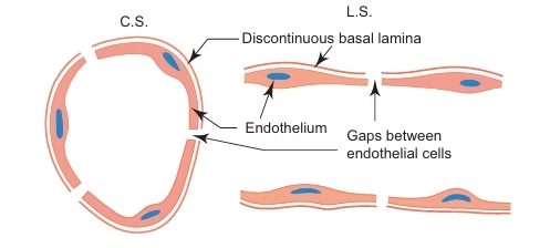
3. Sinusoidal capillary
-
- It is found in liver and haemopoietic organs like red bone marrow and spleen.
- It is a thin walled tortuous blood vessel having a large irregular lumen (30–40 μm).
- Lumen is lined by discontinuous endothelium (the basal lamina is discontinuous).
- There are gaps between the endothelial cells that permit the passage of blood cells and macromolecules.
- Phagocytic cells may be seen in its wall (e.g. Kupffer’s cells in liver).
Veins
Feature
- Veins are thin-walled blood vessels that carry blood from capillaries to heart.
- Large veins are formed by union of smaller veins like tributaries of a river.
- They are often provided with valves which serve to prevent the reflux of the blood.
Structure
The veins are subdivided into the following types based on size.
1. Medium-sized vein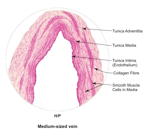
- Medium-sized vein differs from medium-sized artery in having
- A collapsed lumen,
- Thin wall with tunica media containing fewer smooth muscle and less elastic fibres,
- No internal elastic lamina,
- Presence of valves to prevent backflow of blood.
- It is composed of the following three layers:
(a) Tunica intima – It is made of endothelium supported by a thin layer of subendothelium. – It does not have internal elastic lamina.
(b) Tunica media – It is composed of a few circularly arranged smooth muscle fibres embedded in connective tissue predominantly
made of collagen fibres. Elastic fibres are few.
(c) Tunica adventitia – This comprises loose fibroelastic connective tissue carrying vasa vasorum and nerve fibres.
2. Large vein
It is made of the following three layers: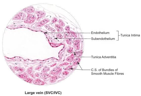
(a) Tunica intima
– This layer is well developed.
– It is formed by endothelium with subendothelial connective tissue.
(b) Tunica media
– It is either thin or absent.
(c) Tunica adventitia
– It is well developed and is the thickest coat.
– It is made of many longitudinal bundles of smooth muscle fibres embedded in connective tissue