Introduction
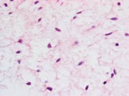
- Connective and other mesenchymal tissues with their stains is one of the four fundamental tissue types in the body, alongside epithelial, muscular, and nervous tissues.
- It is primarily responsible for providing structural and functional support, connecting, and binding various tissues and organs together.
- Connective tissue consists of cells and an extracellular matrix (ECM) that contains fibers (collagen, elastic, reticulin) and ground substance.
- Mesenchymal tissues are a subset of connective tissues derived from mesodermal progenitor cells during embryogenesis.
- These tissues are important for the formation of various body structures, including bones, cartilage, tendons, and muscles.
- Mesenchymal tissues are also involved in tissue repair and regeneration throughout life.

Features of Connective and Mesenchymal Tissues
-
Cells: The cells in connective and mesenchymal tissues include fibroblasts, chondrocytes, osteocytes, adipocytes, and macrophages.
-
Extracellular Matrix (ECM): The ECM consists of fibers (collagen, elastic, reticulin) and ground substance. The ECM’s composition determines the tissue’s mechanical properties, such as rigidity in bones or elasticity in ligaments.
-
Fibers:
-
Collagen fibers: Provide tensile strength and structural integrity.
-
Elastic fibers: Provide elasticity, allowing tissues to stretch and return to their original shape.
-
Reticulin fibers: Form a delicate mesh that supports organs like the liver, spleen, and lymph nodes.
-
Types of Connective Tissues
Connective tissues can be broadly classified into:
-
Loose Connective Tissue: Includes areolar, adipose, and reticular tissue. These tissues provide cushioning and support.
-
Dense Connective Tissue: Contains more collagen fibers and provides strength and flexibility. It includes tendons and ligaments.
-
Specialized Connective Tissues: Such as cartilage, bone, and blood, each with specialized functions.
Staining Methods
Periodic acid-methenamine silver microwave method
Fixative:- 10% neutral buffered formalin is preferred. Mercury-containing fixatives are not recommended.
Sections:- Paraffin-processed tissue cut at 2 μm.
Solutions
- Stock methenamine silver
3% aqueous methenamine 400 ml
5% aqueous silver nitrate 20 ml
Keep refrigerated at 4°C.
- 5% borax (sodium borate) solution
Working methenamine silver solution
Stock methenamine silver 25 ml
Distilled water 25 ml
5% borax 2 ml
- 1% periodic acid solution
0.2% gold chloride solution
1% gold chloride 1 ml
Distilled water 49 ml
- Stock light green solution
Light green SF (yellowish) 1 g
Distilled water 500 ml
Glacial acetic acid 1 ml
- Working light green solution
Light green stock solution 10 ml
Distilled water 50 ml
Method
1. Deparaffinize sections and rehydrate to distilled water.
2. Place sections in 1% periodic acid solution for 15 minutes at room temperature.
3. Rinse in distilled water.
4. Place 5 slides in a plastic Coplin jar containing 50 ml of methenamine working solution. Loosely apply the screw cap and place in the microwave oven, and place a loosely capped plastic Coplin jar containing exactly 50 ml of distilled water in the oven. Microwave (1000 watt) on full power for exactly 70 seconds. Remove both
jars from the oven, mix the solution with a plastic Pasteur pipette and let stand. Check the slides frequently until the desired staining intensity is achieved. This will take approximately 15–20 minutes in a 1000 watt microwave but time calibration may be required.
5. Rinse slides in the heated distilled water.
6. Tone sections in 0.02% gold chloride for 30 seconds.
7. Rinse slides in distilled water.
8. Treat sections with 2% sodium thiosulfate for 1 minute.
9. Wash in tap water.
10. Counterstain in the working light green solution for 11/2 minutes.
11. Dehydrate with two changes each of 95% and absolute alcohol.
12. Clear with xylene and mount with synthetic resin.
Results
Basement membrane Black
Background Green
Van Gieson technique
Sections:- Paraffin. For double embedding in celloidin or low-viscosity nitrocellucose (LVN) sections.
van Gieson solution
Saturated aqueous picric acid solution 50 ml
1% aqueous acid fuchsin solution 9 ml
Distilled water 50 ml
Method
1. Deparaffinize sections and take to water.
2. Stain nuclei by the celestine blue-hematoxylin sequence.
3. Wash in tap water.
4. Differentiate in acid alcohol.
5. Wash well in tap water.
6. Stain in van Gieson solution for 3 minutes.
7. Blot and dehydrate quickly through ascending grades of alcohol.
8. Clear in xylene and mount in permanent mounting medium.
Results
Nuclei Blue/black
Collagen Red
Other tissues Yellow
Masson trichrome technique
Fixation:- Formal sublimate or formal saline.
Sections:- All types.
Preparation of solutions
Solution a
Acid fuchsin 0.5 g
Glacial acetic acid 0.5 ml
Distilled water 100 ml
Solution b
Phosphomolybdic acid 1 g
Distilled water 100 ml
Solution c
Methyl blue 2 g
Glacial acetic acid 2.5 ml
Distilled water 100 ml
Method
1. Deparaffinize sections and take to water.
2. Remove mercury pigment by iodine, sodium thiosulfate sequence.
3. Wash in tap water.
4. Stain nuclei by the celestine blue-hematoxylin method.
5. Differentiate with 1% acid alcohol.
6. Wash well in tap water.
7. Stain in acid fuchsin solution a for 5 minutes.
8. Rinse in distilled water.
9. Treat with phosphomolybdic acid solution b for 5 minutes.
10. Drain.
11. Stain with methyl blue solution c for 2–5 minutes.
12. Rinse in distilled water.
13. Treat with 1% acetic acid for 2 minutes.
14. Dehydrate through ascending grades of alcohol.
15. Clear in xylene, mount in permanent mounting medium.
Results
Nuclei Blue/black
Cytoplasm, muscle, and erythrocytes Red
Collagen Blue
Verhöeff’s method for elastic fibers
Preparation of stain
Solution a
Hematoxylin 5 g
Absolute alcohol 100 ml
Solution b
Ferric chloride 10 g
Distilled water 100 ml
Solution c, Lugol’s iodine
Iodine 1 g
Potassium iodide 2 g
Distilled water 100 ml
Verhöeff’s solution
Solution a 20 ml
Solution b 8 ml
Solution c 8 ml
Add in the above order, mixing between additions.
Method
1. Deparaffinize sections and take to water.
2. Stain in Verhöeff’s solution for15–30 minutes.
3. Rinse in water.
4. Differentiate in 2% aqueous ferric chloride until elastic tissue fibers appear black on a gray background.
5. Rinse in water 6. Rinse in 95% alcohol to remove any staining due to iodine alone.
7. Counterstain as desired, van Gieson is conventional, although eosin may be used.
8. Blot to remove excess stain.
9. Dehydrate rapidly through ascending grades of alcohol.
10. Clear in xylene and mount in permanent mounting medium.
Results
Elastic tissue fibers Black
Other tissues according to counterstain
Aldehyde fuchsin method for elastic fibers
Preparation of staining solution
- Dissolve 1 g of basic fuchsin in 100 ml of 70% ethanol, heat may be used to speed the process.
- After cooling and filtering, add 1 ml of concentrated HCl and 2 ml of paraldehyde.
- Stand at room temperature for 2 days to complete the ripening process which is indicated by a color change from red to purple.
- Ripening time may be reduced by increasing the temperature to 50–60°C.
- The ripened solution should be refrigerated for storage.
- Batches of basic fuchsin suitable for the production of Schiff’s reagent are satisfactory for the preparation of aldehyde fuchsin.
- Paraldehyde may lose some potency upon storage but this may be partially compensated for by the addition of an extra 0.5 ml.
- The staining potential of aldehyde fuchsin is greatest between 2 and 4 days after preparation, but may be adequate for the demonstration of elastic tissue fibers for several weeks if stored at 4°C.
Method
1. Deparaffinize sections and take to water.
2. Oxidize in 1% potassium permanganate for 5 minutes.
3. Rinse in tap water.
4. Remove permanganate staining by treatment with 1% oxalic acid.
5. Rinse in tap water.
6. Rinse in 70% ethanol.
7. Place in sealed container of aldehyde fuchsin for 15 minutes.
8. Rinse well in 70% ethanol.
9. Rinse in tap water.
10. Counterstain as desired, eosin, van Gieson or neutral red are suitable. 11. Dehydrate through ascending grades of alcohol.
12. Clear in xylene and mount in permanent mounting solution.
Results
Elastic tissue fibers blue-purple
Gordon & Sweets’ method for reticular fibers
Preparation of silver solution
- To 5 ml of 10% aqueous silver nitrate solution add concentrated ammonia drop by drop, until the precipitate first formed dissolves, taking care to avoid any excess of ammonia.
- Add 5 ml of 3% sodium hydroxide solution.
- Re-dissolve the precipitate by the addition of concentrated ammonia drop by drop, until the solution retains a trace of opales cence.
- If at this stage any excess of ammonia is present, indicated by the absence of opalescence, add a few drops of 10% silver nitrate solution to produce a light precipitate.
- Make the volume up to 50 ml with distilled water.
- Filter before use. Store in a dark plastic bottle.
Method
1. Deparaffinize sections and take to water.
2. Treat with 1% potassium permanganate solution for 5 minutes.
3. Rinse in tap water.
4. Bleach in 1% oxalic acid solution.
5. Rinse in tap water.
6. Treat with 2.5% iron alum solution for at least 15 minutes.
7. Wash well in several changes of distilled water.
8. Place in a Coplin jar of silver solution for 2 minutes.
9. Rinse in several changes of distilled water.
10. Reduce in 10% aqueous formalin solution for 2 minutes.
11. Rinse in tap water.
12. Tone in 0.2% gold chloride solution for 3 minutes.
13. Rinse in tap water.
14. Treat with 5% sodium thiosulfate solution for 3 minutes.
15. Rinse in tap water.
16. Counterstain as desired.
17. Dehydrate through ascending grades of alcohol.
18. Clear in xylene and mount in permanent mounting medium.
Results
Reticular fibers Black
Nuclei Black or unstained
Gomori’s method for reticular fibers
Preparation of silver solution
- Add 40 ml of 10% silver nitrate solution to 10 ml of 10% potassium hydroxide solution.
- Allow the precipitate to settle and decant the supernatant.
- Wash the precipitate several times with distilled water.
- Add concentrated ammonia drop by drop until the precipitate has just dissolved.
- Add further 10% silver nitrate solution until a little precipitate remains.
- Dilute to 100 ml and filter.
- Store in a dark plastic bottle.
Method
1. Deparaffinize sections and take to water.
2. Treat with 1% potassium permanganate solution for 2 minutes.
3. Rinse in tap water.
4. Bleach in 2% potassium metabisulfate solution.
5. Rinse in tap water.
6. Treat with 2% iron alum for 2 minutes.
7. Wash in several changes of distilled water.
8. Place in Coplin jar of silver solution for 1 minute.
9. Wash in several changes of distilled water.
10. Reduce in 4% aqueous formalin solution for 3 minutes.
11. Rinse in tap water.
12. Tone in 0.2% gold chloride solution for 10 minutes.
13. Rinse in tap water.
14. Treat with 2% potassium metabisulfate solution for 1 minute.
15. Rinse in tap water.
16. Treat with 2% sodium thiosulfate solution for 1 minute.
17. Rinse in tap water.
18. Counterstain as desired, van Gieson or eosin is suitable.
19. Dehydrate through ascending grades of alcohol.
20. Clear in xylene and mount in permanent mounting medium.
Results
Reticular fibers black
Nuclei gray