General feature
- The term connective tissue (CT) is applied to a tissue that fills the interstices between more specialized elements.
- They are predominantly composed of intercellular substance (matrix) secreted by their cells.
- Present in almost every part of the body, it is conspicuous in some regions and scanty in others.
- This kind of CT is referred to as a general CT.
- They are mesodermal in origin.
Classification
Based on the structure and function, it can be classified into:
Embryonic/fetal CT:
-
- Mucoid CT
- Mesenchyme
Connective tissue proper:
-
- Loose CT
- Dense CT—regular and irregular
Specialised CT:
-
- Cartilage
- Bone
- Adipose tissue
- Blood
- Hemolymphatic tissue.
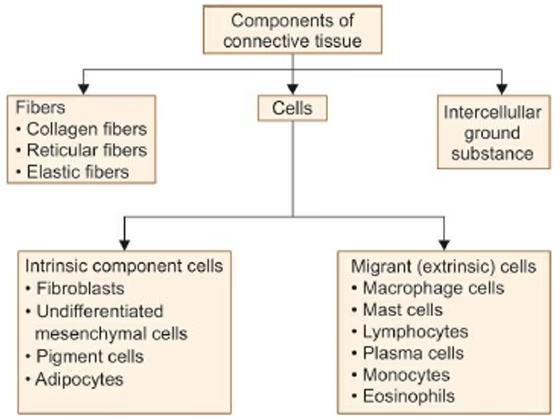
Composition
Connective tissue = Cells + Intercellular matrix (Fibers + Ground substance)

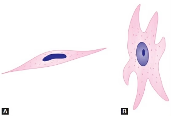
Fibroblasts
Cells
- In tissue sections, the cells appear spindle-shaped, and the nucleus appears to be flattened.
- When seen from the surface, the cells show branching processes.
- The nucleus is large, euchromatic, and has prominent nucleoli.
- They are called fibrocytes when the cytoplasm is scanty, organelles are few, and the nucleus is heterochromatic.
Function
- Fibroblasts become very active when there is a need to lay down collagen fibres; for example, during wound repair, where they form granulation tissue with budding capillaries.
- Fibroblasts are targets of various growth factors which influence cell growth and differentiation.
- In glands and lymphoid tissue, fibroblasts with reticular fibres form a fibrocellular network called reticular tissue.
Pigment Cells
- They are most abundant in CT of the skin, and in the choroid and iris of the eyeball.
- Pigment cells are easily distinguished as they contain brown pigment (melanin) in their cytoplasm and are called melanocytes.
- Melanocytes have rounded cell bodies and long processes.
- They contain melanin granules in a membrane-bound form called melanosomes.

Function
- Variations in the number of pigment cells, and in the amount of pigment in them account for the differences in the skin colour of different races and in different individuals.
- Pigment cells prevent light from reaching other cells. The importance of this function of the eyeball is obvious.
- Pigment cells in the skin protect deeper tissues from the effects of light (especially ultraviolet light). The darker skin of races living in tropical climates is an obvious adaptation for this purpose.
- Albinism is a condition where there is a complete absence of melanin pigment.
Fat Cells (Adipocytes)
- Fat-storing cells are called adipocytes/lipocytes
- Aggregations of fat cells constitute adipose tissue.

- The cytoplasm of the cell forms a thin layer just deep to the plasma membrane.
- The nucleus is pushed against the plasma membrane and is flattened.
- In routine slide preparation, fat is dissolved by organic solvent, thus giving adipocytes a signet ring appearance.
- Adipocytes are incapable of division.
Macrophage Cells
- Macrophage cells of CT are also called histiocytes or clasmatocytes.
- Macrophage cells of CT belong to the mononuclear phagocyte system (MPS).
- They are derived from monocytes, which migrate from the blood.
- Macrophages are usually described as “fixed” when they are attached to fibers.
- Fixed macrophages resemble fibroblasts in appearance.

- They are called motile/nomadic macrophages when they are free, not attached to fibres.
- Free macrophages are rounded and have a regular form.
- However, all macrophages are capable of becoming mobile when suitably stimulated.
- The nuclei of macrophages are smaller, heterochromatic and stain more intensely than those of fibroblasts.
- They are often kidney-shaped.
- Cytoplasm is mildly basophilic. With the electron microscope (EM), the cytoplasm is seen to contain numerous lysosomes that help in “digesting” material phagocytosed by the macrophage.
Mast Cells
- These are large, round or oval cells (mastocytes or histaminocytes)
- The nucleus is small and centrally placed.
- Irregular microvilli (filopodia) are present on the cell surface.
- The distinguishing feature of these cells is the presence of numerous granules in the cytoplasm.

- The granules can be demonstrated with the periodic acid-Schiff (PAS) stain.
- They are most frequently seen around blood vessels and nerves.
- Mast cells are probably related in their origin to basophils of blood.
- The brain and spinal cord are devoid of mast cells; thus, they are protected from the harmful effects of oedema and allergic reactions.
Lymphocytes
- Lymphocytes represent one variety of leukocytes (white blood cells) present in blood.
- Large aggregations of lymphocytes are present in lymphoid tissues.
- They reach CT from these sources, and are especially numerous when the tissue undergoes inflammation.

- Lymphocytes are small with rounded, highly heterochromatic nuclei and a thin rim of cytoplasm.
- They play an important role in the defence of the body against invasion by bacteria and other organisms.
- They can recognise substances that are foreign to the host body and destroy these invaders by producing antibodies against them.
Plasma Cells or Plasmatocytes
- They are round, relatively large cells with basophilic cytoplasm.
- Basophilic cytoplasm is because it has many rough endoplasmic reticulum in it, except for a small region near the nucleus where a well-developed Golgi complex is located.
- The nucleus is spherical and eccentric.

- The chromatin in its nucleus forms four or five clumps near the periphery of the nucleus, thus giving the nucleus a resemblance to a cartwheel appearance.
- Both these features are indicative of the fact that plasma cells are engaged in considerable synthetic activity.
- They produce antibodies that may be discharged locally, may enter the circulation, or may be stored within the cell itself in the form of inclusions called Russell’s bodies.
Fibers of connective tissues
Collagen Fibers
- Collagen fibers are most numerous. They can be classified into various types.
- Reticular fibers were once described as a distinct variety of fibers, but they are now regarded as one variety of collagen fiber.
- With the light microscope, collagen fibers are seen in bundles.
- The bundles may be straight or wavy, depending upon how much they are stretched. The bundles are made up of collections of
individual collagen fibers, which are 1–12 μm in diameter. - The bundles often branch or anastomose with adjacent bundles, but the individual fibers do not branch.
- Unstained collagen fibers in bundles appear white in color with the unaided eye.
- In sections stained with H&E, collagen fibers are stained light pink.
- Collagen fibers are mainly made up of a protein called collagen.
- Protein collagen in turn is made up of molecules of tropocollagen.

Reticular Fibers
- They are much finer and have uneven thickness.
- They form a network (or reticulum) by branching and by anastomosing with each other.
- They do not run in bundles.
- They can be stained specifically by silver impregnation, which renders them black.
- They can, thus, be easily distinguished from type I collagen fibers, which are stained brown.
- Because of their affinity for silver salts, reticular fibers are sometimes called argentophil fibers.
- Reticular fibers provide a supporting network in lymphoid organs, such as the spleen, lymph nodes, and bone marrow; most glands, including the liver and kidneys.
- Reticular fibers form an essential component of all basement membranes.
- They are also found with smooth muscle and nerve fibers.
Elastic Fibers
- Elastic fibers are much fewer than those of collagen. They run singly (not in bundles), branch and anastomose with other fibers.
- Elastic fibers are thinner than those of collagen (0.1–0.2 μm).
- In some situations, elastic fibres are thick (e.g. in the ligamenta flava).
- In other situations (as in the walls of large arteries), they form fenestrated membranes.
- With the EM, each elastic fibre is seen to have a central amorphous core and an outer layer of fibrils.
- Elastic fibers do not stain with the usual stains for collagen. They can be demonstrated by staining with orcein, with aldehyde fuchsin, and by Verhoeff’s method.
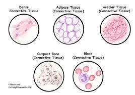
Different forms of connective tissues
Loose Connective/Areolar Tissue
- It is so called because of the loosely arranged collagen fibers in it enclose small spaces (areolae—L. “little open space”) which are filled with interstitial fluid.
- It has abundant ground substance, which makes it soft.
- It has numerous fibroblasts and macrophages along with other types of CT cells scattered between loosely arranged collagen and elastic fibers.
- Its main function is to bind different tissues together.
- Areolar tissue gets distorted easily; hence, it allows the tissue to move freely.
- Examples—endomysium, subperiosteal tissue, lamina propria of gastrointestinal tract (GIT), hypodermis, stroma of glands, and mesentery.
Dense Connective Tissue
- Nuclei of some cells (mainly fibroblasts) are seen between the bundles of collagen.
- Nuclei are elongated in shape (elliptical).

- Ground substance is in a smaller amount.
- In this CT, collagen bundles do not show such a regular arrangement, but interlace in various directions, forming dense irregular tissue.
- Few cells (fibroblast) and less ground substance.
Adipose Tissue
- Adipocytes have single large fat globule, which occupies whole of the cytoplasm.
- In routine sections, the cells appear empty as the fat in them gets dissolved during preparation (treatment with fat solvents, such as xylene or benzene) of the section giving it a honeycomb appearance.
- The cytoplasm of the cell appears as a thin rim around the fat globule.

- The nucleus is flat and lies to one side (eccentric), giving it a signet ring appearance.
- It acts as a storehouse of nutrition, fat being deposited when available in excess, and being removed when deficient in the diet.
- In many situations, fat performs a mechanical function.
- The fat around the kidneys keeps them in position.
- If there is a sudden depletion of this fat, the kidneys may become mobile (floating kidney).