
Working Principle
- Electron microscopes use electron beams instead of light to magnify objects.
- Electrons have a much shorter wavelength than visible light, allowing for much higher magnification (up to 1 million times or more).
-
Electron Beam: The electron gun generates electrons, which are accelerated and focused by electromagnetic lenses. The electron beam interacts with the specimen, either transmitted through (TEM) or reflected off (SEM).
-
Signal Detection: The electrons that interact with the specimen produce various signals, such as secondary electrons or transmitted electrons, which are captured by detectors to form the image.
-
Vacuum: The chamber inside an electron microscope is kept in a vacuum to prevent electrons from scattering due to air particles.
Parts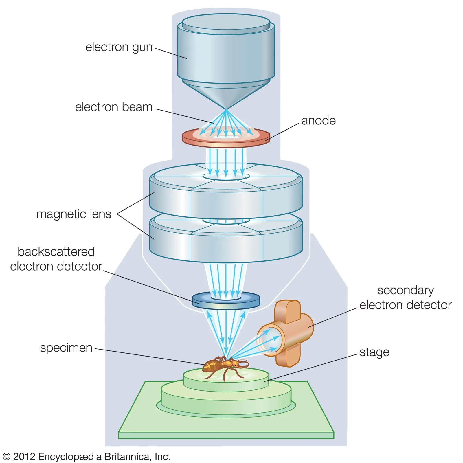
-
Electron Gun: The source of electrons that are accelerated and directed towards the specimen. Typically, a tungsten filament or a field-emission gun is used.
-
Condenser Lens: Focuses the electron beam onto the specimen.
-
Objective Lens: A magnetic lens that focuses the electron beam after it interacts with the sample.
-
Projector Lens: Magnifies the image and projects it onto the viewing screen or detector.
-
Specimen Stage: Holds the specimen in the microscope, often capable of moving along multiple axes.
-
Vacuum System: Electron microscopes operate under a vacuum to avoid electron scattering by air molecules. This includes pumps, chambers, and seals.
-
Viewing Screen or Monitor: Displays the magnified electron image, often via a cathode-ray tube or modern digital screens.
Working Procedure
-
Sample Preparation: Electron microscope samples often need special preparation. For SEM, specimens may be coated with a thin layer of gold or carbon to conduct electrons. For TEM, the sample must be ultra-thin.
-
Place specimen in the chamber: Insert the specimen onto the stage, ensuring it is positioned correctly.
-
Vacuum: The chamber is sealed, and a vacuum is applied.
-
Electron Beam Generation: The electron gun generates a beam that is focused onto the specimen.
-
Interaction with the specimen: The electron beam interacts with the sample, producing secondary or transmitted electrons.
-
Image Formation: These electrons are detected and processed to form a magnified image on a monitor or a photographic plate.
Maintenance
-
Vacuum System: Ensure the vacuum system is regularly checked and leaks are repaired. A strong vacuum is essential for proper operation.
-
Electron Gun: Over time, the electron gun may degrade. Regular inspection and replacement are required to maintain imaging quality.
-
Cleaning: Regularly clean the lenses and specimen holder to prevent contamination.
-
Alignment: Electron microscopes must be carefully aligned to ensure accurate focusing and imaging. Misalignment can reduce resolution.
Applications of Electron Microscope
1. Material Science
-
Nanotechnology: Electron microscopes, especially Transmission Electron Microscopes (TEM) and Scanning Electron Microscopes (SEM), are crucial in the study of nanomaterials. They allow researchers to analyze nanostructures, enabling advances in nanotechnology and materials engineering.
-
Surface Analysis: SEMs are particularly useful for surface morphology analysis of materials like metals, polymers, ceramics, and composites, providing insights into their structure and properties.
2. Biology and Medicine
-
Ultra-Structural Imaging: Electron microscopes are indispensable in studying the ultra-structure of cells and tissues at the subcellular level, including organelles like mitochondria, ribosomes, and lysosomes.
-
Virus and Bacteria Study: TEM and SEM allow researchers to observe viruses, bacteria, and other microorganisms in great detail, which is critical for virology, microbiology, and pathology.
-
Cancer Research: Electron microscopes are used to observe abnormal cell structures in cancerous tissues, helping researchers understand cancer progression at a molecular level.
3. Forensic Science
-
Evidence Examination: In forensic investigations, electron microscopes are used to examine trace evidence such as gunshot residue, fibers, and glass fragments. The detailed images can assist in criminal investigations by providing minute evidence that a light microscope could not detect.
4. Semiconductor and Electronics Industry
-
Chip Fabrication and Quality Control: SEM and TEM are used extensively in the semiconductor industry for inspecting integrated circuits (ICs) and chips at the nanometer scale. They help identify defects in materials and ensure the quality of electronic components.
-
Surface and Interface Analysis: Electron microscopes are used to analyze the surfaces and interfaces of materials in the development of new electronic devices, allowing for the optimization of material properties and performance.
5. Environmental Science
-
Pollution Analysis: Electron microscopes are used to study particulate matter in the atmosphere or sediments, offering a detailed understanding of environmental pollutants and their effects. They can detect airborne pollutants such as soot, dust, and bacteria.
-
Microplastics Study: They are employed in environmental research to identify and analyze microplastics in water, soil, and other environmental samples, which is critical for assessing their impact on ecosystems and human health.
6. Archaeology and Art Conservation
-
Cultural Heritage Preservation: Electron microscopes are used to analyze ancient artifacts, paintings, and other cultural objects. They help conservators study materials used in the objects (e.g., pigments, metals, ceramics) at the microscopic level, aiding in preservation techniques.
-
Fossil Analysis: Electron microscopes are used in paleontology to study fossils, allowing scientists to examine the intricate details of fossilized tissues and structures.
7. Education and Research
-
High-Resolution Imaging: Electron microscopes are invaluable in academic and industrial research for obtaining high-resolution images of materials, biological samples, and nanoscale structures, facilitating breakthroughs in various scientific fields.
-
Training for Advanced Research: They are essential tools for training researchers and students in highly specialized scientific fields, particularly in nanotechnology, materials science, and molecular biology.
8. Geology
-
Mineral Identification: Electron microscopes can be used to study the structure of minerals at the atomic and molecular levels, helping geologists identify and analyze the composition of minerals and rock samples.