Introduction

History of Electron Microscope
- The electron microscope was developed in 1931 by Ernst Ruska and Max Knoll.
- Ernst Ruska later received the Nobel Prize in Physics (1986) for his contribution.
- The first commercial electron microscope became available in the late 1930s.
- Electron microscopy greatly improved the understanding of cellular and microbial structures.
Principle
The electron microscope works on the principle that:
Electrons have a much shorter wavelength than visible light, allowing higher resolution and magnification.
In an electron microscope:
- A beam of electrons is generated from an electron gun.
- The electron beam is focused by electromagnetic lenses.
- Electrons interact with the specimen.
- The image formed is magnified and viewed on a fluorescent screen or computer monitor.
Basic Components of Electron Microscope
1. Electron Gun
The electron gun is the source of electrons in the microscope. It produces a beam of high-speed electrons required for imaging.
Components
- Tungsten filament or field emission source
- Cathode
- Anode
Functions
- Generates electrons by heating the filament.
- Accelerates electrons toward the specimen using high voltage.
- Produces a narrow electron beam.
Importance
- It is the primary source of illumination in the electron microscope.
2. Electromagnetic Lenses
Electron microscopes use electromagnetic lenses instead of glass lenses to focus electrons.
Types
- Condenser lens
- Objective lens
- Projector lens
Functions
- Focus and control the electron beam.
- Magnify the image produced from the specimen.
- Improve image clarity and resolution.
Principle
- Magnetic fields bend and focus the path of electrons.
3. Condenser Lens
The condenser lens is located below the electron gun.
Functions
- Concentrates the electron beam onto the specimen.
- Controls beam intensity and diameter.
- Provides uniform illumination.
Importance
- Proper focusing improves image quality and contrast.
4. Specimen Holder (Stage)
The specimen holder or stage supports the specimen during examination.
Functions
- Holds the specimen securely.
- Allows movement and positioning of the sample.
- Maintains specimen stability.
Features
- Made of metal grids or special holders.
- In TEM, ultra-thin sections are mounted on copper grids.
5. Objective Lens
The objective lens is the most important lens in the electron microscope.
Functions
- Produces the first magnified image of the specimen.
- Determines the resolution of the microscope.
- Focuses transmitted or reflected electrons.
Importance
- Responsible for fine structural details and image sharpness.
6. Projector Lens
The projector lens further enlarges the image produced by the objective lens.
Functions
- Provides additional magnification.
- Projects the image onto the viewing screen or camera.
Importance
- Helps achieve extremely high magnification levels.
7. Vacuum Chamber
Electron microscopes require a high vacuum system because electrons cannot travel effectively through air.
Functions
- Removes air molecules from the chamber.
- Prevents scattering of electrons.
- Maintains a clear electron pathway.
Importance
- Essential for proper electron movement and image formation.
8. Fluorescent Screen
The fluorescent screen converts electron signals into visible images.
Functions
- Produces visible images when struck by electrons.
- Allows direct observation of the specimen.
Importance
- Used for image viewing and focusing.
9. Camera and Image Detector
Modern electron microscopes contain digital cameras and detectors.
Functions
- Capture and store images.
- Provide digital image analysis.
- Improve image recording quality.
Importance
- Useful for research, diagnosis, and documentation.
10. Aperture
An aperture is a small opening that controls the electron beam.
Functions
- Regulates electron flow.
- Improves image contrast.
- Reduces unwanted scattered electrons.
Importance
- Enhances image sharpness and clarity.
11. Cooling System
Electron microscopes generate heat during operation.
Functions
- Prevents overheating.
- Maintains instrument stability.
Importance
- Protects sensitive components from damage.
12. Power Supply System
The microscope requires a stable high-voltage power supply.
Functions
- Provides electrical energy to the electron gun and lenses.
- Controls electron acceleration.
Importance
- Ensures stable and efficient microscope functioning.
Types of Electron Microscopes
1. Transmission Electron Microscope (TEM)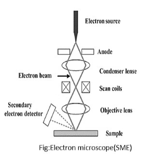
Working Principle:
- In TEM, a high-energy electron beam is produced by an electron gun, typically using a tungsten filament or a field emission gun. The beam is then condensed and focused onto the specimen by the condenser lens system.
- As the electrons pass through the specimen, they interact with the atoms and can be transmitted or scattered. The extent of scattering depends on the density and composition of the material. The objective lens then focuses the transmitted electrons to form an image on a fluorescent screen or a digital camera.
Key Features:
- Resolution: TEM can achieve resolutions better than 1 nm, enabling the observation of individual atoms in materials.
- Contrast Mechanisms: Contrast in TEM images arises from differences in electron density, the thickness of the specimen, and the atomic number of the elements present.
Typical Applications:
- Analysis of cell organelles, such as the mitochondria and endoplasmic reticulum.
- Structural analysis of viruses, proteins, and nanoscale materials.
- Characterization of crystal structures and defects in materials science.
Components:
- Electron Gun: Generates the electron beam, often using a tungsten filament heated to produce thermionic emission or a field emission source for higher brightness and coherence.
- Condenser Lenses: Focus the electron beam onto the specimen; multiple lenses can be used to achieve a small spot size.
- Specimen Stage: Holds the specimen and allows for precise movement (e.g., tilting and rotation) during imaging.
- Objective Lens: Primary lens that magnifies the image created by the transmitted electrons.
- Intermediate and Projector Lenses: Further magnify and project the image onto a screen or detector.
- Detector System: Includes photographic films, fluorescent screens, or digital cameras for image capture.
2. Scanning Electron Microscope (SEM)
Working Principle:
- SEM operates by scanning a finely focused electron beam across the surface of a specimen. The electrons interact with the atoms in the specimen, producing secondary electrons (for surface imaging), backscattered electrons (for compositional contrast), and characteristic X-rays (for elemental analysis).
- The emitted signals are collected by detectors and processed to create a high-resolution, three-dimensional image of the specimen surface.
Key Features:
- Depth of Field: SEM provides a greater field depth than optical microscopy, allowing for clear imaging of the specimen’s surface morphology.
- Three-Dimensional Imaging: By scanning the surface and compiling data, SEM can produce three-dimensional representations of the specimen.
Typical Applications:
- Surface morphology analysis of biological specimens, such as tissues, cells, and bacteria.
- Examination of materials, including fractures, coatings, and composites.
- Semiconductor and nanotechnology research for characterizing microstructures.
Components:
- Electron Gun: Generates and focuses the electron beam; often employs a tungsten filament or a field emission source.
- Condenser Lenses: Focuses the electron beam onto the sample surface.
- Scanning Coils: Direct the beam across the specimen in a raster pattern, allowing for scanning.
- Detectors: Collect emitted signals:
- Secondary Electron Detector: Most commonly used for imaging surface topography.
- Backscattered Electron Detector: Provides information about elemental composition and contrast based on atomic number.
- X-ray Detector (EDX): For elemental analysis, detects X-rays emitted during electron interactions.
Difference Between TEM and SEM
| Feature | TEM | SEM |
|---|---|---|
| Full Form | Transmission Electron Microscope | Scanning Electron Microscope |
| Image | Internal structure | Surface structure |
| Specimen | Ultra-thin section | Whole specimen |
| Resolution | Very high | Moderate |
| Appearance | 2D image | 3D image |
| Magnification | Higher | Lower than TEM |
| Electron Path | Through specimen | Over specimen surface |
Allied Techniques for Electron Microscopy
1. Cryo-Electron Microscopy (Cryo-EM)
Cryo-EM is a variant of TEM that enables the imaging of biological specimens in their native hydrated states by rapidly freezing them. This avoids the artifacts that may arise from chemical fixation and dehydration.
Advantages:
- Preserving biological samples in their near-native state allows for better structural analysis of macromolecules.
- No need for staining, which can introduce artifacts or alter structures.
Applications:
- Structural biology to study protein complexes, ribosomes, and membrane proteins.
- Visualization of cellular structures, such as membranes and cytoskeleton components.
2. Electron Tomography
Electron tomography is a technique to obtain specimens’ three-dimensional (3D) structures from a series of 2D images taken at different angles.
Process:
- Multiple images of the same specimen are collected at different tilt angles.
- Computational algorithms reconstruct these images into a 3D representation, allowing for detailed spatial analysis of complex structures.
Applications:
- Studying the 3D architecture of cells, organelles, and other biological structures at high resolution.
- Analysis of material properties and defects.
3. Energy-dispersive X-ray Spectroscopy (EDX/EDS)
EDX is an analytical technique commonly used in conjunction with SEM and TEM to determine the elemental composition of a specimen.
Process:
- When the electron beam interacts with the specimen, it excites the atoms, causing them to emit characteristic X-rays.
- The EDX detector measures the energy and intensity of the emitted X-rays, allowing for identifying elements present in the sample.
Applications:
- Elemental analysis of biological tissues, minerals, and materials.
- Characterization of nanomaterials and coatings.
4. Immunoelectron Microscopy
This technique combines immunolabeling with electron microscopy to visualize specific proteins or antigens in cellular contexts.
Process:
- Specimens are treated with antibodies that bind to the target antigen.
- The antibodies are conjugated to electron-dense markers, such as colloidal gold.
- The sample is imaged using TEM, revealing the location of the labeled antigens.
Applications:
- Localizing proteins within cells, such as receptors, enzymes, and structural proteins.
- Studying virus-host interactions by identifying viral proteins in infected cells.
5. Focused Ion Beam (FIB) Microscopy
FIB is often used with SEM for sample preparation and imaging, allowing for precise milling and cross-sectional imaging.
Process:
- A focused beam of ions (commonly gallium) is used to sputter away material from the surface of a specimen, creating a cross-section or shaping the sample for analysis.
Applications:
- Preparing thin samples for TEM by milling the surface to a suitable thickness.
- Creating patterns on materials for microfabrication and nanotechnology applications.
Advantages
- Extremely high magnification
- Very high resolution
- Detailed ultrastructural study
- Visualization of viruses and organelles
- Three-dimensional imaging possible with SEM
- Important in research and diagnosis
Limitations
- Very expensive
- Requires skilled operators
- Large instrument size
- Complex specimen preparation
- Specimens must be dead
- Cannot observe living cells directly
- Requires vacuum environment
Applications
In Medicine
- Diagnosis of kidney diseases
- Cancer research
- Study of muscle disorders
- Viral identification
In Microbiology
- Study of bacteria and viruses
- Analysis of microbial ultrastructure
In Biochemistry
- Protein and enzyme structure analysis
- Molecular studies
In Nanotechnology
- Nanoparticle characterization
- Surface analysis
In Material Science
- Metal and alloy examination
- Semiconductor analysis
In Forensic Science
- Gunshot residue analysis
- Hair and fiber examination
Recent Advances in Electron Microscopy
- Cryo-electron microscopy (Cryo-EM)
- Digital imaging systems
- Atomic-level imaging
- Automated image analysis
- High-resolution 3D reconstruction