Introduction
- Entamoeba gingivalis is a protozoan parasite that primarily resides in the oral cavity.
- It was the first amoeba discovered in humans, identified in 1849 by Friedrich Lösch. Unlike other species of the Entamoeba genus (e.g., Entamoeba histolytica), E. gingivalis is not associated with the gastrointestinal tract but is instead implicated in oral diseases, including periodontal disease.
- While it has traditionally been considered a commensal organism, increasing evidence suggests it may contribute to pathological processes, particularly in periodontal inflammation and tissue destruction.
Geographical Distribution
Entamoeba gingivalis is found worldwide, with no specific geographical restrictions. Its prevalence varies depending on oral hygiene practices, age, and the presence of periodontal disease.
-
- Higher prevalence:
- Individuals with poor oral hygiene.
- Patients with periodontal disease, gingivitis, or other oral infections.
- Lower prevalence:
- Populations with good dental care and oral hygiene practices.
- Higher prevalence:
Habitat
-
- E. gingivalis primarily resides in the oral cavity, particularly in the:
- Gingival crevices.
- Periodontal pockets.
- Tonsillar crypts.
- Dental plaques.
- The parasite feeds on epithelial cells, bacteria, leukocytes, and other debris found in these regions, especially in areas with active inflammation or infection.
- E. gingivalis primarily resides in the oral cavity, particularly in the:
Morphology
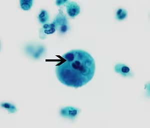
Unlike other Entamoeba species, E. gingivalis exists only as a trophozoite and does not form cysts.
-
- Trophozoite:
- Shape: Irregular, amoeboid.
- Size: 10–20 µm in diameter.
- Structure:
- A single nucleus with a central karyosome and peripheral chromatin.
- Cytoplasm may contain ingested material, such as leukocytes and bacteria (erythrophagocytosis is not observed).
- Highly motile due to the presence of pseudopodia.
- Trophozoite:
Life Cycle
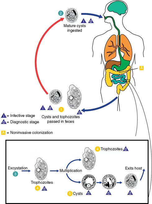
The life cycle of E. gingivalis is simple and involves only the trophozoite stage.
-
- Colonization:
- Trophozoites inhabit the gingival crevices and periodontal pockets of the oral cavity.
- Replication:
- The trophozoites reproduce asexually by binary fission.
- Transmission:
- Directly transmitted from person to person via saliva or contaminated objects.
- Colonization:
Mode of Transmission
-
- gingivalis is transmitted primarily through:
-
- Saliva exchange:
- Kissing or sharing utensils, toothbrushes, or other oral items.
- Contact with oral secretions:
- Close personal contact or indirect transmission through contaminated dental instruments.
- Association with dental procedures:
- Invasive dental treatments may facilitate its spread.
- Saliva exchange:
Unlike E. histolytica, E. gingivalis is not transmitted through the fecal-oral route since it lacks a cyst stage.
Incubation Time
There is no defined incubation period for E. gingivalis as it is not always pathogenic. Its presence in the oral cavity may persist for extended periods without causing noticeable symptoms unless oral hygiene deteriorates or periodontal disease develops.
Pathogenesis
Entamoeba gingivalis is traditionally considered a commensal organism; however, its role in oral disease pathogenesis is increasingly recognized.
-
- Mechanisms of Pathogenesis:
- Adhesion to tissues: E. gingivalis adheres to oral epithelial cells, particularly in areas with inflammation.
- Phagocytosis: Engulfs leukocytes, bacteria, and cellular debris, potentially exacerbating tissue damage.
- Enzymatic activity: Secretes proteolytic enzymes may degrade host tissues and extracellular matrix components, contributing to periodontal disease progression.
- Associated Conditions:
- Periodontal disease: Frequently found in patients with gingivitis or periodontitis.
- Halitosis: Its presence has been linked to bad breath due to tissue breakdown and bacterial activity.
- Immune Response:
- E. gingivalis may evade the immune system by engulfing immune cells, such as neutrophils.
- Chronic inflammation in periodontal tissues is associated with its presence.
- Mechanisms of Pathogenesis:
Laboratory Diagnosis
Diagnosis of E. gingivalis involves detecting trophozoites in oral samples.
-
- Microscopy:
- Direct microscopic examination of dental plaques, gingival scrapings, or periodontal pocket material.
- Trophozoites are identified by their characteristic morphology and motility.
- Staining Methods:
- Trichrome or Giemsa staining enhances the visualization of trophozoites.
- Molecular Techniques:
- Polymerase chain reaction (PCR) can detect E. gingivalis DNA in oral samples with high sensitivity and specificity.
- Culture:
- In vitro cultivation is rarely performed but can be used for research purposes.
- Microscopy:
Treatment
Treatment of E. gingivalis infections focuses on eliminating the parasite and addressing underlying oral conditions, such as periodontal disease.
-
- Antiparasitic Therapy:
- Metronidazole: Effective against E. gingivalis trophozoites. Typically prescribed in combination with dental treatments.
- Chlorhexidine mouthwash: Reduces microbial load and may help control parasite numbers.
- Dental Care:
- Professional dental cleaning to remove plaques and calculus.
- Periodontal therapy for patients with gum disease.
- Oral Hygiene:
- Regular brushing, flossing, and use of antiseptic mouthwashes to maintain a healthy oral environment and prevent recolonization.
- Antiparasitic Therapy:
Prevention
-
- Maintain Good Oral Hygiene:
- Brush and floss regularly to minimize plaque buildup.
- Avoid Sharing Oral Items:
- Avoid sharing utensils, toothbrushes, or dental appliances.
- Regular Dental Checkups:
- Routine professional cleaning and assessment can reduce the risk of colonization.
- Sterilization of Dental Instruments:
- Proper sterilization practices in dental clinics prevent cross-contamination.
- Maintain Good Oral Hygiene: