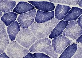
- Enzyme histochemistry is a method for localizing and visualizing enzymatic activities within tissue sections.
- Here’s a detailed overview of the principles, materials, reagents, procedures, and expected results for demonstrating the enzymatic activities of phosphatase, dehydrogenase, oxidase, and peroxidase in histochemistry.
Phosphatase Histochemistry
- Phosphatase enzymes, including acid and alkaline phosphatases, play critical roles in metabolic processes like bone mineralization and lysosomal activity.
- In histopathology, demonstrating phosphatase activity can help identify tissues with high lysosomal activity or areas of bone remodelling.
Reagents and Substrates
- Alkaline Phosphatase:
- Substrate: Naphthol AS-BI phosphate, which releases naphthol on dephosphorylation.
- Diazonium salt: Fast Blue BB or Fast Red TR forms an insoluble azo dye bound to naphthol, producing a visible color.
- Acid Phosphatase:
- Substrate: Naphthol AS-BI phosphate is similar to alkaline phosphatase but used in an acidic environment.
- Diazonium salt: Fast Garnet or Fast Red Violet is commonly used to yield a reddish reaction product.
- Buffers:
- Alkaline Buffer: Tris buffer, pH 9-10, helps maintain enzyme activity for alkaline phosphatase.
- Acidic Buffer: Acetate buffer, pH 4.5-5, stabilizes acid phosphatase activity.
- Fixatives: Acetone or very brief formalin fixation preserves enzyme function. Heavy fixation can denature enzymes, resulting in a weak or absent reaction.
Procedure Steps
- Slide Preparation: Tissue should be fresh or snap-frozen to retain enzyme activity.
- Fixation: Sections are fixed briefly to maintain cellular morphology without inactivating phosphatases.
- Incubation in Substrate Solution:
- For alkaline phosphatase: Sections are incubated in the alkaline naphthol AS-BI solution with Fast Blue BB at 37°C for up to 30 minutes.
- For acid phosphatase: Sections are incubated in the acidic naphthol AS-BI solution with Fast Garnet at 37°C.
- Stop Reaction: The sections are rinsed with distilled water to halt enzyme activity, ensuring well-defined end products.
- Counterstaining: Sections can be lightly counterstained with hematoxylin to enhance nuclear detail without masking enzyme activity.
Results Interpretation
- Alkaline Phosphatase: Positive staining is observed in osteoblasts, liver cells, kidney tubules, and placenta, with blue to purple coloration at activity sites.
- Acid Phosphatase: Found within lysosomes, macrophages, and certain neurons. Red to violet granules indicate a positive reaction, especially in sites of high lysosomal activity.
Dehydrogenase Histochemistry
Dehydrogenases, such as succinate dehydrogenase and lactate dehydrogenase, are crucial for cellular respiration and energy metabolism. Their activity is demonstrated by reducing tetrazolium salts to colored formazan compounds within tissues.
Reagents and Substrates
- Substrates:
- Succinate for Succinate Dehydrogenase (SDH): Specific for mitochondrial respiratory activity.
- Lactate for Lactate Dehydrogenase (LDH): Reflects glycolytic pathway activity.
- Hydrogen Acceptors: Nitro-blue tetrazolium (NBT) or tetrazolium chloride acts as an electron acceptor and forms a colored formazan precipitate upon reduction.
- Buffers: A phosphate buffer, pH 7.4, is generally used to maintain a stable environment.
- Fixative: Frozen sections without fixation or acetone fixation for best enzyme retention, as fixation can inactivate dehydrogenases.
Procedure Steps
- Slide Preparation: Use 10–12 µm cryostat sections, maintaining cold conditions to preserve enzymes.
- Incubation with Substrate and Tetrazolium Salt: Sections are incubated at 37°C in a solution containing the substrate (e.g., succinate or lactate) and the tetrazolium salt in a phosphate buffer.
- Formazan Precipitate Formation: Enzyme activity reduces the tetrazolium salt to a visible formazan compound.
- Rinsing and Stopping Reaction: Distilled water halts the reaction, preserving the colored end product.
- Counterstaining (Optional): Light counterstain may be applied, though typically avoided to preserve contrast.
Results Interpretation
- Succinate Dehydrogenase: High activity in mitochondria-rich cells, such as cardiac and skeletal muscle fibers, with a deep blue or purple formazan precipitate. Helps visualize mitochondrial abnormalities in myopathies.
- Lactate Dehydrogenase: Indicates cytoplasmic activity, with a reddish-blue deposit in cells relying on anaerobic glycolysis, like muscle fibers under hypoxia.
Oxidase Histochemistry
Oxidases, particularly cytochrome oxidase, are vital for mitochondrial function and cellular respiration. This stain is valuable in tissues like the brain, where cytochrome oxidase activity varies among neuron populations.
Reagents and Substrates
- Substrates: Diaminobenzidine (DAB) or tetrazolium salts are common, reacting with oxidase activity to yield a colored product.
- Hydrogen Peroxide: Often included to serve as an electron donor in reactions involving DAB.
- Buffers: Phosphate buffer, pH 7.0–7.4, maintains enzyme stability.
- Fixative: Light acetone or formalin fixation may be used sparingly, as excessive fixation impairs oxidase function.
Procedure Steps
- Slide Preparation: Tissue sections are cut frozen and often used without fixation.
- Substrate Reaction: Sections are incubated in a solution of DAB and hydrogen peroxide at room temperature.
- Oxidation Reaction: Oxidase enzymes catalyze oxidation of DAB, forming a brown precipitate.
- Rinse and Mount: Wash with distilled water to halt the reaction and prevent diffusion of the end product.
Results Interpretation
- Cytochrome Oxidase: Positive activity results in a brown precipitate in neurons, especially in brain areas like the cortex and hippocampus, where it aids in identifying neuronal metabolic activity and detecting mitochondrial disorders.
Peroxidase Histochemistry
Peroxidases, like neutrophils and macrophages, such as myeloperoxidase and horseradish peroxidase, demonstrate cells involved in inflammatory responses. Peroxidase activity is also exploited in immunohistochemistry.
Reagents and Substrates
- Substrate: Diaminobenzidine (DAB) or aminoethylcarbazole (AEC) as chromogenic substrates for peroxidase.
- Hydrogen Peroxide: Reacts with DAB to produce a colored precipitate.
- Buffers: Phosphate buffer, pH 7.4.
- Fixative: Light formalin fixation if required, though often used in fresh-frozen sections for immunohistochemical applications.
Procedure Steps
- Slide Preparation: Sections are frozen or lightly fixed, especially for immunohistochemical applications.
- Incubation with Substrate and Hydrogen Peroxide: Sections are treated with DAB and hydrogen peroxide in phosphate buffer.
- Color Development: Peroxidase catalyzes the breakdown of hydrogen peroxide, oxidizing DAB to form a visible brown product.
- Rinsing and Counterstaining: Rinse with distilled water to halt the reaction, and sections can be counterstained lightly to enhance cellular detail.
Results Interpretation
- Myeloperoxidase: Found in neutrophils and macrophages, producing a brown deposit in areas of high inflammatory cell activity.
- Horseradish Peroxidase (HRP): Frequently used in immunohistochemistry to visualize antibody binding, with a distinct brown or red coloration when DAB or AEC is used.