AIM: Gross Examination of Histopathological Specimens
Principle
- The principle of gross examination of histopathology involves the visual and tactile assessment of tissue specimens before they undergo microscopic examination.
- The primary goal is to document the specimen’s size, shape, color, texture, and any abnormalities that may provide clues to the underlying pathology.
- The findings from the gross examination help in:
-
-
Selecting representative tissue areas for microscopic analysis.
-
Guiding the processing of the specimen (such as sectioning and embedding).
-
Documenting key features that may be important for diagnosis or surgical planning.
-
Material Requirements
-
Tissue Specimens
-
Dissecting Tools: Sharp instruments such as scalpels, scissors, forceps, and knives are used for handling, dissecting, and sectioning the tissue.
-
Fixative
-
Measuring Instruments
-
Recording Materials
-
Specimen Containers
-
Surgical Markers
-
Personal Protective Equipment
Sample Labelling
-
Patient Information
-
Specimen Details
-
Date and Time
-
Special Instructions
-
Barcode or QR Code
Grossing Procedure
-
Specimen Reception: When a specimen is received, check for completeness and proper labeling. Ensure that all accompanying paperwork or forms are present.
-
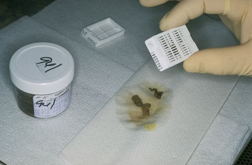
External Examination: Visual inspection of the specimen is carried out first, noting the size, shape, color, and texture. Any obvious abnormalities like tumors, cysts, or hemorrhages should be documented.
-
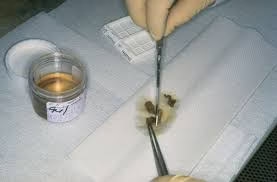
Dissection: The specimen is cut into smaller pieces to evaluate its internal characteristics. Care should be taken to avoid damaging delicate tissue.
-
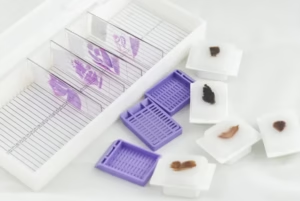
Selection of Representative Samples: Based on the visual examination, select representative tissue portions that will give a comprehensive view of the pathology. These include areas of abnormal tissue, tumor margins, or any lesions.
-
Photographic Documentation: Photographs of the specimen may be taken for record-keeping or for reference in future diagnoses.
-
Measuring: The specimen is measured using a ruler or calipers. Record the dimensions in millimeters or centimeters.
-
Detailed Recording: Document all findings in a structured format, describing the specimen’s features in detail, including any abnormal findings.
-
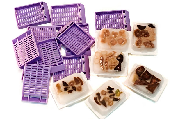
Preservation: Once the specimen has been dissected and the necessary areas for microscopic examination are selected, it is placed in formalin for fixation before further processing.
Collection of Samples
-
Biopsy: A small portion of the suspected tissue is removed using a needle, endoscope, or surgical technique. Proper aseptic technique must be followed to avoid contamination.
-
Surgical Resection: Larger pieces of tissue are removed during surgery. These should be carefully handled and stored in formalin or another appropriate fixative.
-
Autopsy: Tissue samples are taken from various organs post-mortem, ensuring the proper sequence and selection of organs for pathological analysis.
Fixative
Fixatives are chemicals used to preserve tissue samples and prevent decomposition or alteration before microscopic analysis. The most commonly used fixative is formalin (10% neutral-buffered formalin), which preserves tissue morphology and cellular structure. Other fixatives include:
-
Alcohol-based Fixatives: Often used for preserving cytology specimens.
-
Bouin’s Solution: Used for certain types of tissues that require better preservation of finer details (e.g., connective tissue).
-
Zinc-based Fixatives: These may be used for immunohistochemistry.
Transport of Sample
-
Use of Appropriate Containers: Tissue should be placed in leak-proof containers that are filled with an adequate amount of fixative (such as formalin) to fully immerse the specimen.
-
Temperature Control: Specimens should be transported at room temperature. Avoid freezing, which can damage cellular structures.
-
Timeliness: Transport the specimen as soon as possible to minimise degradation. Ideally, the sample should reach the lab within a few hours, especially if it is large or from a vital organ.
-
Labelling: Ensure the specimen is labelled clearly, including patient information, specimen details, and any relevant clinical history or special instructions.
Care and Maintenance
-
Handling: Handle specimens gently to avoid crushing, tearing, or distorting the tissue. Always use sharp, clean tools for dissection.
-
Clean Tools: Dissecting instruments should be cleaned and sterilised between uses to prevent cross-contamination between specimens.
-
Storage: Specimens should be stored in fixative (commonly formalin) until further processing. Ensure that containers are securely closed to prevent leakage.
-
Workplace Hygiene: Maintain a clean laboratory environment. Regularly disinfect surfaces and equipment.
-
Equipment Maintenance: Ensure that equipment such as microscopes, refrigerators, and tissue processors are properly calibrated and maintained to ensure reliable performance.
-
Personal Safety: Lab workers should wear gloves, lab coats, and safety glasses to protect themselves from hazardous materials such as formalin and other chemicals.