Joints, or articulations, are connections between two or more bones that allow for movement and flexibility in the skeletal system. They play a crucial role in the body’s mobility, enabling various activities, from simple walking to complex motions like dancing or playing sports. Joints can vary significantly in their structure, function, and range of motion, adapting to the mechanical needs of the body.
Classification of Joints
-
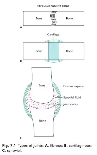
Structural Classification

This classification is based on the type of connective tissue that binds the bones and whether a joint cavity is present.
- Fibrous Joints:
- Characteristics: Connected by dense connective tissue, generally immovable.
- Subtypes:
- Sutures:
- Location: Found between the bones of the skull.
- Function: Allow for minimal movement; provide strength and stability to the skull.
- Syndesmoses:
- Location: Between the tibia and fibula (interosseous membrane).
- Function: Slightly movable, allowing some degree of flexibility.
- Gomphoses:
- Location: Tooth sockets in the mandible and maxilla.
- Function: Immovable; provide stability to teeth.
- Sutures:
- Cartilaginous Joints:
- Characteristics: Bones are united by cartilage; no joint cavity is present.
- Subtypes:
- Synchondroses:
- Location: Epiphyseal plates in growing long bones, costal cartilage connecting ribs to the sternum.
- Function: Allow for growth; provide flexibility.
- Symphyses:
- Location: Pubic symphysis, intervertebral discs.
- Function: Slightly movable; provides strength and absorbs shock.
- Synchondroses:
- Synovial Joints:
- Characteristics: Most common and movable joints are characterized by a joint cavity.
- Subtypes:
- Hinge Joints:
- Location: Elbow, knee, interphalangeal joints.
- Function: Permit flexion and extension.
- Ball-and-Socket Joints:
- Location: Shoulder (glenohumeral joint), hip (acetabulofemoral joint).
- Function: Allow movement in multiple planes and rotation.
- Pivot Joints:
- Location: Atlantoaxial joint (between the first and second cervical vertebrae).
- Function: Allow rotational movement.
- Condyloid Joints:
- Location: Wrist (radiocarpal joint), metacarpophalangeal joints.
- Function: Permit flexion, extension, and some rotation.
- Saddle Joints:
- Location: Carpometacarpal joint of the thumb.
- Function: Allow for opposition and grasping.
- Plane Joints:
- Location: Intercarpal joints of the wrist.
- Function: Allow gliding movements.
- Hinge Joints:
-
Functional Classification
This classification is based on the degree of movement allowed by the joint.
- Synarthroses: Immovable joints. Examples include sutures in the skull.
- Amphiarthroses: Slightly movable joints. Examples include the pubic symphysis and intervertebral joints.
- Diarthroses: Freely movable joints, all of which are synovial joints. They allow a wide range of movement and include most limb joints.
Structure of Joints
-
Components of Synovial Joints
Understanding the structure of synovial joints is crucial due to their complexity and the variety of movements they facilitate.
- Articular Cartilage:
- Description: Smooth, hyaline cartilage covering the ends of bones at the joint.
- Function: Reduces friction during movement and absorbs shock.
- Joint Capsule:
- Description: A fibrous sleeve that surrounds the joint.
- Components:
- Outer Fibrous Layer: Provides strength and stability to the joint.
- Inner Synovial Membrane: Produces synovial fluid and lines the joint capsule.
- Synovial Fluid:
- Description: A viscous fluid that fills the joint cavity.
- Function: Lubricates the joint, nourishes articular cartilage, and reduces friction.
- Ligaments:
- Description: Strong bands of dense connective tissue connecting bones to bones.
- Function: Stabilize the joint and restrict excessive movement.
- Tendons:
- Description: Connect muscle to bone.
- Function: Facilitate movement by transmitting force from muscles to bones.
- Bursae:
- Description: Small, fluid-filled sacs located around joints.
- Function: Reduce friction between moving structures, such as tendons and bones.
-
Additional Structures in Some Joints
- Menisci: C-shaped cartilaginous structures in the knee joint that provide cushioning and stability.
- Fat Pads: Adipose tissue that cushions and fills spaces within the joint.
- Labrum: A fibrocartilaginous rim around the socket of ball-and-socket joints, deepening the joint and providing stability (e.g., glenoid labrum in the shoulder).
Functions of Joints
- Facilitating Movement:
- Joints are the points of articulation where bones interact, allowing for various movements. Different types of joints enable specific types of motion, such as flexion, extension, rotation, and gliding.
- Providing Stability:
- Joints maintain structural integrity and stability while allowing movement. Ligaments, tendons, and the joint capsule support and prevent dislocations.
- Absorbing Shock:
- The combination of articular cartilage and synovial fluid helps to absorb impact during activities such as walking, running, or jumping, protecting the bones from injury.
- Permitting Flexibility:
- Joints provide the flexibility necessary for a wide range of activities. The design of various joint types allows for both stability and mobility, accommodating different movements required in daily life.
- Supporting Weight:
- Joints, particularly in the lower body, are critical for bearing weight and maintaining posture. The arrangement of joints helps distribute the body’s weight, providing balance and stability during movement.
- Facilitating Body Posture:
- Joints work in concert with muscles to maintain body posture and alignment. They allow subtle adjustments to keep the body stable during various activities.
Synovial Fluid
Synovial fluid is a viscous, lubricating fluid found within the cavities of synovial joints. It is critical in joint health and function, facilitating smooth movement between articulating bones.
Composition
Synovial fluid is composed of several key components:
- Hyaluronic Acid:
- A glycosaminoglycan that provides viscosity and elasticity, allowing for lubrication and shock absorption.
- Lubricin:
- A glycoprotein that further enhances lubrication and reduces friction between the articular cartilage surfaces.
- Electrolytes:
- Like plasma, synovial fluid contains electrolytes like sodium, potassium, and chloride, which help maintain osmotic balance.
- Nutrients:
- Provides essential nutrients to the avascular articular cartilage, including glucose and oxygen.
- Cells:
- It contains synoviocytes (cells of the synovial membrane) and immune cells (macrophages and lymphocytes) that play a role in joint health and immune response.
Functions
Synovial fluid has several important functions in the body:
- Lubrication:
- Reduces friction between articular surfaces during joint movement, enhancing smooth motion.
- Shock Absorption:
- Distributes load and absorbs impact, protecting the cartilage and underlying bones during activities like walking and running.
- Nutrient Distribution:
- It supplies nutrients and oxygen to the avascular articular cartilage, which lacks blood supply.
- Waste Removal:
- Assists in removing metabolic waste products from the cartilage and joint space.
- Immune Function:
- It contains immune cells that help protect the joint from infection and inflammation.
Production
- Source: Synovial fluid is produced by the joint capsule’s synovial membrane.
- Regulation: Production is influenced by joint activity, with increased movement generally leading to higher fluid production to accommodate the needs of the joint.
Clinical Relevance
Understanding synovial fluid is crucial in various clinical contexts:
- Joint Disorders:
- Abnormalities in synovial fluid can indicate joint disorders. For instance:
- Osteoarthritis: Often leads to decreased viscosity and increased inflammatory components in synovial fluid.
- Rheumatoid Arthritis: May show increased volume, higher cell counts, and inflammatory markers.
- Abnormalities in synovial fluid can indicate joint disorders. For instance:
- Joint Aspiration (Arthrocentesis):
- This procedure involves extracting synovial fluid from a joint for diagnostic purposes. Analysis can help diagnose conditions such as:
- Infections: Elevated white blood cell counts may indicate septic arthritis.
- Crystals: The presence of crystals can indicate gout or pseudogout.
- This procedure involves extracting synovial fluid from a joint for diagnostic purposes. Analysis can help diagnose conditions such as:
- Visco-supplementation:
- Hyaluronic acid injections are used as a treatment for osteoarthritis to improve lubrication and relieve pain by enhancing the properties of synovial fluid.
- Synovitis:
- Inflammation of the synovial membrane can lead to increased production of synovial fluid, causing joint swelling and pain.
- Diagnostic Tests:
- Laboratory analysis of synovial fluid can reveal important information about joint health, including cell count, viscosity, and the presence of crystals or pathogens.