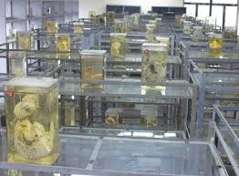
- Museum techniques in histopathology are meticulous procedures used to preserve, display, and maintain biological specimens for long-term use in educational, research, and historical contexts.
- These techniques cover various specimen types and involve wet and dry preservation methods.
- Below is an in-depth exploration of the full process, including variations, specialized techniques, and best practices.
Process of Museum Techniques
-
Specimen Selection and Collection
Specimen selection is critical as only well-preserved, structurally intact samples will offer value in a museum setting. Selected specimens should exhibit anatomical or pathological features of interest, such as diseased tissues, developmental anomalies, or unique structures. Selection may include normal and pathological samples to show comparative anatomy for educational specimens.
- Acquisition: Specimens are collected either fresh (immediately post-surgery or autopsy) or as previously fixed tissues.
- Initial Cleaning: If necessary, the specimen is washed in a saline solution to remove blood or debris.
-
Dissection and Preparation of the Specimen
- Trimming: Specimens are trimmed to highlight specific features or structures. For example, an organ may be cut longitudinally to expose interior pathology.
- In Situ, Placement: Some specimens, such as limbs or organs, may be posed in a natural anatomical position to aid educational display.
- Injection Techniques: Vascular systems are sometimes injected with colored latex or resin to enhance the visibility of arteries, veins, or other vascular structures, which is particularly useful for teaching anatomy.
-
Fixation
Fixation is essential to prevent autolysis and putrefaction of tissues, preserving cellular and structural integrity for long-term storage. The type of fixative used depends on the specimen type, the desired final appearance, and whether the specimen will undergo further processing.
- Formalin (10%): The most commonly used fixative, providing excellent structural integrity and tissue firmness preservation.
- Kaiserling’s Solution I: Ideal for color preservation. Specimens are first fixed in Kaiserling Solution I for several days, then transferred to Kaiserling Solution II or III.
- Alcohol: Ethanol or isopropanol may be used for delicate tissues or specimens undergoing staining. Alcohol-based fixatives offer excellent structural preservation but may cause tissue shrinkage if used for prolonged periods.
-
Dehydration and Clearing
This step is necessary for certain specimens embedded in resins or specimens undergoing plastination or dry preservation. In dehydration, water is gradually replaced by a dehydrating agent like alcohol.
- Alcohol Series: Specimens are passed through a graded series of alcohol solutions, starting at 70% and increasing to 100%.
- Clearing Agents: After dehydration, xylene or other organic solvents may be used to prepare the tissue for embedding in resins or for plastination.
-
Preservation Techniques
Wet Preservation Techniques
Wet preservation involves storing specimens in a fluid medium that prevents decomposition, discoloration, and bacterial growth.
- Formalin-Based Solutions: A mixture of 10% formalin diluted to 4% is used for permanent preservation. It provides stability and resists microbial growth.
- Kaiserling Solution II or III: After initial fixation in Kaiserling Solution I, this fluid enhances color retention for organs like the lungs or kidney, where natural color aids in understanding pathology.
- Glycerin Solutions: Glycerin, sometimes mixed with alcohol, maintains tissue flexibility and softness. Glycerin-based solutions are particularly useful for fatty or delicate tissues.
- Other Solutions: Buffered formalin, alcohol-formalin mixtures, or other clear solutions may provide a transparent, non-cloudy appearance for high-visibility displays.
Dry Preservation Techniques
Certain specimens are processed as dry mounts for long-term, durable storage without fluid.
- Air Drying and Freezing: Specimens are thoroughly fixed and dehydrated before drying in a controlled environment or freezer. This technique is often used for bones, soft tissues, and whole organs.
- Freeze-Drying: A more advanced method, freeze-drying involves freezing the specimen and then reducing surrounding pressure to allow the sublimation of ice. This preserves tissue structure without the shrinkage seen in air drying.
- Plastination: This advanced process replaces water and lipids with curable polymers like silicone, epoxy, or polyester. Plastinated specimens are durable, non-toxic, and retain flexibility, making them suitable for hands-on learning.
- Procedure: The specimen is fixed, dehydrated, cleared, and immersed in the polymer solution under vacuum conditions. The polymer infiltrates the tissue, and the specimen is cured under light or heat to harden.
Corrosion Casting
In corrosion casting, the vascular or ductal systems are filled with a colored resin, which hardens and is freed from surrounding tissue by corrosive agents like potassium hydroxide.
- Applications: Ideal for displaying vascular networks or other intricate tubular systems within organs.
- Procedure: The vascular system is filled with resin and allowed to harden. The surrounding tissue is dissolved, leaving a cast of the vascular structure.
-
Mounting, Display, and Labelling
Mounting and Displaying Specimens
- Containers: Transparent glass or acrylic containers are used for wet-preserved specimens. The container size should be proportional to the specimen to limit movement and fluid evaporation.
- Suspension Techniques: Specimens may be suspended on transparent supports, placed on stands, or fixed using clear nylon threads or fine metal supports.
- Sealing: Containers are sealed with rubber gaskets, silicone, or wax to prevent leakage, evaporation, and contamination. Screw caps or clamps ensure a tight seal.
Labeling
Proper labeling is essential for educational use and long-term cataloging.
- Information: Labels should include the specimen name, type, key anatomical or pathological features, date of preparation, preservation method, and, if possible, donor or source information.
- Positioning: Labels should be positioned so they do not obstruct the view of the specimen but are easily readable. Waterproof ink or engraved labels are ideal for durability.
-
Staining and Coloring (Optional)
Certain stains and dyes may enhance tissue visual contrast, highlighting structures or pathological changes.
- Common Stains: Carmine, eosin, and Prussian blue are commonly used for enhancing the visual clarity of organs, blood vessels, or other tissue types.
- Procedure: Staining is performed post-fixation, where specimens are briefly immersed in the dye, rinsed, and then transferred to a preservation solution.
Maintenance of Museum Specimens
Museum specimens require periodic maintenance to ensure preservation quality over time.
- Regular Solution Replacement: Preservation fluids, especially formalin and glycerin solutions, should be replaced periodically to avoid discoloration, microbial growth, or evaporation.
- Inspection: Specimens should be inspected regularly for signs of mold, leakage, or fluid cloudiness, indicating bacterial contamination or chemical breakdown.
- Environmental Control: Specimens should be stored in low-light, controlled environments to reduce the effects of UV light, temperature fluctuations, and humidity on specimen integrity.
Advanced Museum Techniques and Innovations
High-Fidelity Replication
With advances in 3D scanning and printing technology, high-resolution models of specimens can now be created for study without risking the integrity of rare or fragile specimens. This allows students and researchers to handle and explore highly detailed replicas of anatomical structures.
Digital Museum Collections
High-resolution imaging and digital cataloging systems have allowed museums to create extensive digital collections. These digital repositories can store images and detailed data on each specimen, enabling researchers and educators worldwide to access information remotely.
Challenges and Considerations
- Microbial Contamination: Solutions like glycerin or formalin require antimicrobial additives or periodic replacement to prevent fungal or bacterial growth.
- Color Fading: Natural and artificial light can degrade colors in wet and dry specimens over time. Storage in low-light areas or containers with UV-protective coatings can mitigate this issue.
- Structural Degradation: Over time, some specimens may shrink, become brittle, or lose clarity due to the chemical composition of the preservation fluids. Monitoring and proper chemical maintenance are essential.