
Principle
- Blood smear preparation is used to examine blood cells under a microscope.
- It allows for the evaluation of the morphology of blood cells, identification of abnormalities, and determination of cell counts in cases of hematological disorders.
- Preparing a blood smear involves spreading a thin layer of blood on a microscope slide, which is then stained to allow for better visualization of cell structures.
हिंदी नोट्स के लिए यहां क्लिक करें
Material Requirements
-
Microscope slides: Clean, grease-free slides.
-
Cover slips: Optional, depending on the need for oil immersion.
-
Capillary tubes or lancets: For blood collection.
-
Pipettes or droppers: For transferring blood to the slide.
-
Staining reagents: Depending on the method, common stains include:
-
Wright’s stain, Giemsa stain, or Romanowsky stains.
-
Fixatives (like methanol) may be required in some methods.
-
-
Distilled water: For rinsing the slides after staining.
-
Forceps: For handling slides.
Procedure
-
Blood Collection:
-
A small drop of blood is obtained using a lancet or capillary tube.
-
It is essential to use a fresh sample for optimal results.

-
-
Placing the Blood on the Slide:
-
Place a small drop of blood (approximately 2–3 mm) on one end of the clean slide.
-
-
Spreading the Blood:
-
Hold another slide at a 30–45° angle to the slide with the blood drop.
-
Use the angled slide to spread the blood drop across the surface of the slide in a smooth, even manner by pulling the blood along the slide.
-
-
Air-Drying:
-
Allow the slide to air dry completely to avoid distortion during staining.
-
-
Fixation:
-
If necessary, fix the smear by dipping it in methanol or air-drying.
-
-
Staining:
-
Wright’s or Giemsa Staining Method:
-
Place a drop of Wright’s or Giemsa stain on the dried smear and allow it to spread evenly.
-
Let the stain sit for a few minutes (usually 1–2 minutes).
-
Wash the slide gently with distilled water and let it air dry again.
-
-
Romanowsky Staining:
-
After fixation, use Romanowsky stains for differential staining of blood components.
-
-
-
Examination:
-
Examine the slide under a microscope using the oil immersion objective for detailed observation of blood cells.
-
हिंदी नोट्स के लिए यहां क्लिक करें
Haematological Methods of Staining Blood Smears
Romanowsky Staining:
Principle:
- Romanowsky staining is a method that uses a mixture of acidic and basic dyes to differentiate various cellular components in blood smears.
- The basic dye (methylene blue) stains acidic components like the nuclei of cells, while the acidic dye (eosin) stains the cytoplasm and red blood cells (RBCs).
- This dual staining method enables the visualization of different cell types in blood, especially white blood cells (WBCs), with their distinct morphology.
Procedure:
-
Blood Smear Preparation:
-
First, prepare a blood smear by placing a drop of blood on a clean glass slide and spreading it thinly with another slide at a 30–45° angle.
-
Allow the smear to air dry.
-
-
Fixation:
-
The smear is fixed by immersing the slide in methanol for a few minutes to preserve the cell structures.
-
-
Staining:
-
Apply the Romanowsky stain (typically Wright’s or Leishman) to the dried, fixed smear.
-
Leave the stain on the slide for 2-3 minutes.
-
Add a small amount of buffer solution or distilled water to dilute the stain and gently mix it for 3-5 minutes.
-
-
Washing and Drying:
-
After the staining process, rinse the slide with distilled water to remove excess stain.
-
Let the slide air dry completely before examination under the microscope.
-
Uses:
-
Romanowsky staining is widely used in routine blood smears to differentiate between various types of blood cells (RBCs, WBCs, platelets).
-
It is also used for identifying cell abnormalities, such as changes in the shape, size, and number of cells, which are useful in diagnosing conditions like leukemia, anemia, and infections.
-
This method is particularly useful for differential leukocyte counts and for detecting blood parasites, such as Plasmodium (malaria) and Babesia.
Leishman Staining:
Principle:
- Leishman stain is a Romanowsky stain composed of eosin and methylene blue.
- It is used to differentiate and examine blood cells, especially white blood cells.
- Leishman stain selectively stains cellular structures based on their affinity for acidic or basic dyes.
- The methylene blue (basic dye) stains the nucleus of the cells, while eosin (acidic dye) stains the cytoplasm.
- Leishman staining provides excellent results for examining granular leukocytes, as it stains the granules in neutrophils, eosinophils, and basophils.
- This makes it a valuable technique for investigating blood in clinical haematology.
Procedure:
-
Blood Smear Preparation:
-
Place a small drop of blood on a clean glass slide and spread it thinly using another slide at an appropriate angle.
-
Allow the smear to air dry.
-
-
Fixation:
-
Fix the smear by immersing it in methanol for 3–5 minutes, preserving the cell structures.
-
-
Staining:
-
Apply Leishman stain to the smear and let it sit for 2-3 minutes.
-
Afterward, dilute the stain with buffer solution or distilled water in a 1:2 ratio and mix for 3-5 minutes.
-
-
Washing and Drying:
-
Rinse the slide with distilled water to remove excess stain.
-
Let the slide air dry completely before examining it under the microscope.
-
Uses:
-
Leishman staining is commonly used to identify and differentiate white blood cells, especially for distinguishing between neutrophils, eosinophils, and basophils.
-
It is particularly useful in diagnosing and monitoring conditions such as leukemia, malaria, and other blood infections.
-
It is also beneficial for examining the morphology of the nucleus and cytoplasm of blood cells, which helps identify diseases like parasitic infections and hematologic malignancies.
-
Leishman stain is frequently used for detecting malaria parasites in blood samples, as it highlights the presence of Plasmodium in red blood cells.
हिंदी नोट्स के लिए यहां क्लिक करें
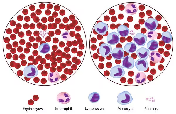
Morphology of Normal Blood Cells
-
Red Blood Cells (RBCs) – Erythrocytes:
-
Shape: Biconcave disc-shaped cells.
-
Size: 7-8 µm in diameter.
-
Color: Pinkish with a pale center due to the absence of a nucleus.
-
Function: Transport of oxygen and carbon dioxide.

-
-
White Blood Cells (WBCs) – Leukocytes:
-
Types:
-
Neutrophils:
-
Size: 12-15 µm.
-
Nucleus: Multi-lobed.
-
Cytoplasm: Pale, with fine granules.
-
Function: Phagocytosis of bacteria.
-
-
Lymphocytes:
-
Size: 7-9 µm.
-
Nucleus: Large, round, occupying most of the cell.
-
Cytoplasm: Scanty and pale blue.
-
Function: Immunity, producing antibodies.
-
-
Monocytes:
-
Size: 12-17 µm.
-
Nucleus: Large, kidney-shaped or bean-shaped.
-
Cytoplasm: Abundant, pale blue.
-
Function: Phagocytosis, antigen presentation.
-
-
Eosinophils:
-
Size: 12-17 µm.
-
Nucleus: Bi-lobed.
-
Cytoplasm: Bright orange-pink, contains large granules.
-
Function: Defense against parasites and allergic reactions.
-
-
Basophils:
-
Size: 12-17 µm.
-
Nucleus: Bi-lobed.
-
Cytoplasm: Large, purple granules.
-
Function: Release histamine and heparin in allergic reactions.
-
-
-
-
Platelets – Thrombocytes:
-
Size: 2-4 µm in diameter.
-
Shape: Small, disk-shaped fragments.
-
Color: Blue cytoplasm with purple granules.
-
Function: Blood clotting, helping in hemostasis.
-