
Introduction
- A peripheral blood smear is a vital tool for evaluating the morphology and characteristics of blood cells.
- The three major cell types found in blood are red blood cells (RBCs), white blood cells (WBCs), and platelets.
- Their identification based on morphology under the microscope helps diagnose various haematological and systemic diseases.
- Below is a more detailed description of these cells, including size, shape, structure, and cytoplasmic characteristics.
- Blood is a specialized connective tissue that circulates throughout the body.
- It consists of plasma and formed elements.
- The formed elements include:
- Red blood cells (RBCs)
- White blood cells (WBCs)
- Platelets
- Study of blood cell morphology is important because identification of normal cells helps detect many blood disorders.
- Blood cell morphology is usually studied on a peripheral blood smear stained by Romanowsky stains such as Leishman stain, Wright stain, or Giemsa stain.
Red Blood Cells 
Morphological Characteristics:
- Shape: Red blood cells are biconcave discs, allowing flexibility and a large surface area-to-volume ratio. This shape facilitates gas exchange and passage through narrow capillaries.
- Size: RBCs have a diameter of 7-8 µm.
- Colour: They stain a pale pink to red on Romanowsky stains (like Wright’s or Giemsa’s) because of their haemoglobin content.
- Central Pallor: RBCs exhibit a pale central area representing their thinner, biconcave center. This central pallor occupies about one-third of the RBC’s diameter.
- Nucleus: Mature RBCs are anucleate (they do not contain a nucleus), characteristic of their fully differentiated state.
- Function: RBCs transport oxygen from the lungs to tissues and carry carbon dioxide back to the lungs for exhalation.
Identification:
- Under the microscope, RBCs appear round, biconcave discs that stain evenly with a distinct central pale area.
- They should not have inclusions, and their size and shape should be relatively uniform.
Common Observations in a Healthy Smear:
- RBCs should be evenly distributed without significant clumping or overlapping.
- In normal smears, the RBCs should exhibit minimal variations in size (anisocytosis) or shape (poikilocytosis).
White Blood Cells
White blood cells, or leukocytes, are critical in immune responses. They are divided into two main categories: granulocytes (containing cytoplasmic granules) and agranulocytes (lacking prominent granules).
Granulocytes
These cells contain prominent cytoplasmic granules that stain different colours with Romanowsky stains. Granulocytes include neutrophils, eosinophils, and basophils.
Neutrophils (Polymorphonuclear Neutrophils or PMNs)
- Shape: Neutrophils have a multilobed nucleus (typically 3-5 lobes connected by thin chromatin strands). This gives them the name “polymorphonuclear.”
- Size: They measure about 12-15 µm in diameter.
- Cytoplasm: The cytoplasm contains fine, neutral-staining granules, which are faint and difficult to see with standard staining. These granules contain enzymes like myeloperoxidase, which aid in bacterial killing.
- Colour: Neutrophils exhibit a pale pink or lilac cytoplasm with fine granules.
- Function: Neutrophils are the body’s first defence in bacterial infections, responsible for phagocytosis and killing pathogens.
Identification:
- Neutrophils are easily recognized by their multilobed nucleus and faint, granular cytoplasm.
- They account for most WBCs (about 60-70% in a normal differential count).
Eosinophils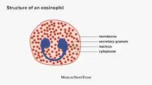
- Shape: Eosinophils have a bilobed nucleus (two lobes connected by a thin chromatin strand).
- Size: They measure about 12-15 µm in diameter.
- Cytoplasm: The cytoplasm is packed with large, red-orange granules. These granules are rich in proteins like major basic protein and eosinophil peroxidase, which play a role in combating parasites.
- Colour: The large acidophilic granules are bright orange-red with Romanowsky stains.
- Function: Eosinophils are primarily involved in responses to parasitic infections and allergic reactions.
Identification:
- Eosinophils are easily recognized by their large, bright red granules and bilobed nuclei.
- They make up about 2-4% of the WBC count.
Basophils
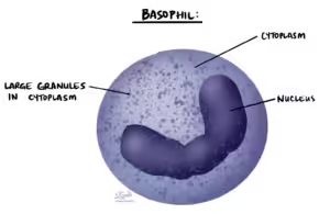
- Shape: Basophils have a bilobed or S-shaped nucleus, although the granules often obscure the nucleus.
- Size: They measure about 10-14 µm in diameter.
- Cytoplasm: The cytoplasm is filled with large dark blue-purple granules, which often overlap and obscure the nucleus.
- Colour: The large basophilic granules stain dark purple or blue-black.
- Function: Basophils are involved in allergic reactions and release histamine and heparin during immune responses.
Identification:
- Basophils are identified by their large, dark blue granules, which often obscure the nucleus.
- They are the least common type of WBC, constituting less than 1% of the WBC count.
Agranulocytes
Agranulocytes do not contain visible cytoplasmic granules. This group includes lymphocytes and monocytes.
Lymphocytes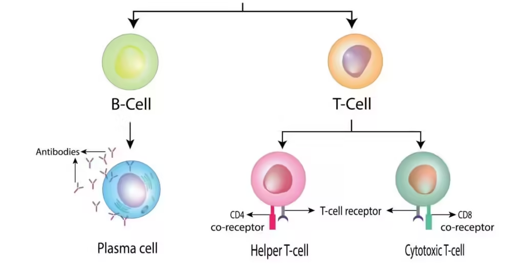
- Shape: Lymphocytes are round with a large, round nucleus that almost fills the entire cell.
- Size: Small lymphocytes are 7-10 µm, while large lymphocytes can be 10-15 µm.
- Cytoplasm: The cytoplasm is scant and stains a pale blue. The cytoplasm forms a narrow rim around the nucleus in small lymphocytes, whereas large lymphocytes have more abundant cytoplasm.
- Nucleus: The nucleus is typically round and deeply staining (dark purple).
- Function: Lymphocytes are key players in the immune response, with B cells involved in antibody production and T cells in cell-mediated immunity.
Identification:
- Lymphocytes have a large, dense nucleus with a small amount of blue cytoplasm.
- They represent 20-40% of WBCs.
Monocytes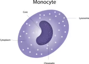
- Shape: Monocytes have an irregular, indented, or kidney-shaped nucleus.
- Size: They are the largest WBCs, measuring 15-20 µm in diameter.
- Cytoplasm: The cytoplasm is abundant and stains grey-blue, often containing fine granules and vacuoles.
- Nucleus: The nucleus is often described as horseshoe-shaped or folded.
- Function: Monocytes are responsible for phagocytosis and mature into macrophages when they enter tissues and ingest foreign substances and dead cells.
Identification:
- Monocytes are larger than other WBCs and have a characteristic kidney-shaped nucleus.
- They make up about 2-8% of the WBC count.
Platelets 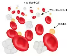
Morphological Characteristics:
- Shape: Platelets are small, disc-shaped cell fragments.
- Size: They measure 2-4 µm in diameter, making them the smallest of the blood elements.
- Colour: Platelets stain a pale blue with scattered purple granules.
- Nucleus: Platelets are anucleate (they do not have a nucleus) because they are fragments of megakaryocytes.
- Function: Platelets are essential for blood clotting and wound healing. They aggregate at the site of vascular injury and release factors that promote clot formation.
Identification:
- Platelets appear as small, irregularly shaped, purple-staining granules in the background of the blood smear, often clustered together.
- They can be found scattered between the larger RBCs and WBCs.
Table of Normal Blood Cell Morphology
| Cell Type | Size | Nucleus | Cytoplasm | Main Identification |
|---|---|---|---|---|
| RBC | 7–8 µm | Absent | Pink with central pallor | Biconcave disc |
| Neutrophil | 10–14 µm | Multilobed | Fine granules | Segmented nucleus |
| Eosinophil | 10–14 µm | Bilobed | Orange-red granules | Large granules |
| Basophil | 10–12 µm | Bilobed | Deep blue granules | Granules obscure nucleus |
| Lymphocyte | 7–10 µm | Round dense | Thin blue rim | Large nucleus |
| Monocyte | 12–20 µm | Kidney-shaped | Gray-blue | Largest WBC |
| Platelet | 2–4 µm | Absent | Purple granules | Small fragments |
Clinical Importance
- Study of normal blood cell morphology is very important because it helps in recognizing abnormal blood cells during microscopic examination.
- Identification of normal red blood cells, white blood cells, and platelets is the first step in hematological diagnosis.
- Changes in blood cell size, shape, color, or number often indicate underlying disease.
Clinical Importance of Red Blood Cell Morphology
- Variation in RBC size helps diagnose anemia.
- Small RBCs (microcytes) are seen in iron deficiency anemia.
- Large RBCs (macrocytes) are seen in vitamin B12 or folate deficiency.
- Variation in RBC shape may indicate hereditary disorders or hemolytic anemia.
- Pale RBCs suggest reduced hemoglobin content.
- Presence of abnormal inclusions may indicate specific blood diseases.
Clinical Importance of White Blood Cell Morphology
- Increase in neutrophils usually suggests bacterial infection.
- Increase in eosinophils is commonly seen in allergy and parasitic infection.
- Increased lymphocytes may occur in viral infections.
- Increased monocytes may be seen in chronic infections.
- Presence of immature white cells may indicate leukemia.
- Nuclear changes in WBCs help in identifying bone marrow disorders.
Clinical Importance of Platelet Morphology
- Low platelet number may lead to bleeding disorders.
- Large platelets may suggest increased platelet production.
- Platelet clumping may interfere with platelet count.
- Abnormal platelet morphology may indicate platelet function disorders.
Importance in Peripheral Blood Smear Examination
- Peripheral smear examination provides direct visual information about blood cells.
- It helps confirm automated blood count findings.
- It is essential in diagnosis of anemia, leukemia, infections, and clotting disorders.
Importance in Laboratory Practice
- Correct identification of normal cells prevents reporting errors.
- It improves accuracy in differential leukocyte count.
- It is essential for students, laboratory technicians, and clinicians during routine hematology work.