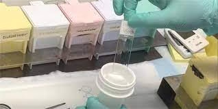
Introduction
- A histological fixative is a chemical substance used to preserve biological tissues from decay, maintaining the structure of cells and tissues for microscopic examination.
- Fixatives stabilise the tissue, preventing autolysis (self-digestion) and putrefaction (decay due to bacteria).
- They preserve tissue by killing microorganisms, stopping enzymatic activities, or cross-linking proteins, ensuring the tissue retains its original form for analysis.

Properties of Histological Fixatives:
- Preservation: Fixatives stabilize the biological structure, maintaining the integrity of tissues and cells as close as possible to their living state.
- Penetration: Effective fixatives should penetrate the tissue quickly and uniformly.
- Cross-linking or Precipitation: Many fixatives act by cross-linking proteins or precipitating components to prevent degradation and maintain tissue morphology.
- Hardening: Fixatives may harden the tissue to facilitate easier sectioning for microscopy.
- Compatibility: The fixative should be compatible with subsequent staining techniques to not interfere with visualising specific cellular or tissue components.
- Isotonicity: Ideally, the fixative should not cause swelling or shrinkage of cells, maintaining cellular morphology.
Type of material obtained in laboratory
The human tissue comes from the surgery, and the autopsy room from surgery, two types of tissue are obtained.
- As a biopsy- A small piece of lesions or tumour is sent for diagnosis before the final removal of the lesion or the tumour (Incisional biopsy).

- If the whole tumour or lesion is sent for examination and diagnosis by the pathologist, it is called an excisional biopsy.
- Tissues from the autopsy are sent to study disease and its course for advancing medicine.
Types of Histological Preparation
The histological specimen can be prepared as 1. Whole mount 2. Sections 3. Smears.
- Whole mounts- These are preparation for entire animals, e.g. fungi, and parasites. These preparations should be no more than 0.2-0.5 mm in thickness.
- Sections- The majority of the preparations in histology are sections. The tissue is cut into about 3-5 mm thick pieces processed, and 5 microns thick sections are cut on a microtome.
- Smears are made from blood, bone marrow, or pleural or ascitic fluid. These are immediately fixed in alcohol to present the cellular structures and are then stained.
Responsibility of a technician
- Specimen preservation.
- Specimen labelling, logging and identification.
- Preparation of the specimen to facilitate their gross and microscopy.
- Record keeping.
Type of fixative
- Simple fixative
- Compound fixative
Simple fixative
A simple fixative consists of a chemical substance used for tissue fixation instead of compound fixatives that combine several chemicals. These fixatives are straightforward to use and provide specific preservation based on the nature of the tissue and the fixative’s properties.
-
Formaldehyde (Formalin):
- Description: A 4% formaldehyde solution (10% neutral buffered formalin) is the most widely used simple fixative. Formaldehyde forms cross-links between proteins, primarily through amino acids like lysine.
- Properties: It preserves the overall structure of the tissue well, though it may cause some shrinkage. It penetrates tissues rapidly and allows for long-term storage.
- Usage: Common in general histology for light microscopy, preserving tissues for routine examination.
-
Glutaraldehyde:
- Description: A simple fixative that is a stronger aldehyde than formaldehyde, commonly used in electron microscopy.
- Properties: It produces extensive cross-linking between proteins, resulting in excellent preservation of cellular ultrastructure, but it penetrates tissue more slowly than formaldehyde.
- Usage: Ideal for electron microscopy due to its ability to preserve fine structural details.
-
Ethanol:
- Description: Ethanol is a simple alcohol-based fixative precipitating proteins and nucleic acids.
- Properties: It is a dehydrating fixative that removes water from tissues, preserving cellular components but causing significant shrinkage and hardening.
- Usage: Often used in cytology for smears and in preparing samples for DNA or RNA analysis.
-
Osmium Tetroxide:
- Description: Osmium tetroxide is a simple fixative used mainly in electron microscopy.
- Properties: It reacts with lipids and helps preserve membranes and other lipid-containing structures, giving excellent contrast in electron micrographs. However, it is toxic and expensive.
- Usage: Often used as a secondary fixative following aldehyde fixation in electron microscopy.
Advantages of Simple Fixatives:
-
- Easy to prepare and use.
- Good for specific types of tissue preservation based on the chosen fixative.
- Many are compatible with routine staining techniques.
Disadvantages of Simple Fixatives:
-
- They may not provide the best results for all tissue types, requiring additional fixatives or secondary treatments for optimal preservation.
Compound fixative
A compound fixative combines two or more chemical substances better to preserve tissue than a single (simple) fixative. By combining different fixatives, compound fixatives leverage the strengths of each component, improving the fixation of various cellular components.
-
Bouin’s Solution:
- Components: Picric acid, formaldehyde, and acetic acid.
- Properties:
- Picric acid enhances the preservation of soft tissues and cellular details.
- Formaldehyde cross-links proteins, maintaining the structure of the tissue.
- Acetic acid helps preserve nucleic acids and prevents tissue shrinkage.
- Usage: Commonly used for soft tissues, particularly in the endocrine system and for embryos. It is also used in plant tissue fixation but may interfere with some staining methods due to its strong yellow colour from picric acid.
-
Carnoy’s Solution:
- Components: Ethanol, chloroform, and acetic acid.
- Properties:
- Ethanol is a dehydrating agent that precipitates proteins.
- Chloroform aids in tissue penetration.
- Acetic acid preserves nucleic acids and prevents excessive hardening or shrinkage.
- Usage: Excellent for fixing chromosomes, used frequently in cytology, and ideal for rapid fixation of small tissues.
-
Zenker’s Fixative:
- Components: Mercuric chloride, potassium dichromate, acetic acid, and sodium sulfate.
- Properties:
- Mercuric chloride preserves fine nuclear details and enhances staining.
- Potassium dichromate stabilizes cytoplasmic structures, especially mitochondria.
- Acetic acid ensures good nuclear preservation.
- Usage: Often used for bone marrow and blood tissues. It provides excellent detail for hematopoietic tissues but requires careful handling due to mercury toxicity.
-
Helly’s Fixative:
- Components: Mercuric chloride, potassium dichromate, formaldehyde.
- Properties: Combines the penetration and preservation capabilities of mercuric chloride and potassium dichromate with formaldehyde’s protein cross-linking properties.
- Usage: Used primarily for preserving blood cells and tissues containing blood, particularly for histological studies of the spleen and bone marrow.
-
Zamboni’s Fixative (Buffered Picric Acid-Formaldehyde):
- Components: Picric acid and formaldehyde.
- Properties:
- Formaldehyde cross-links proteins for tissue stability.
- Picric acid improves tissue penetration and preserves fine cellular details.
- Usage: Useful for general tissue fixation and immunohistochemistry because of its compatibility with many staining procedures.
Advantages of Compound Fixatives:
-
- Balanced Preservation: Compound fixatives often provide more balanced tissue preservation by combining the effects of different chemicals (e.g., protein cross-linking, nucleic acid preservation, lipid stabilization).
- Improved Detail: They can enhance the visualization of specific cellular components like the nucleus, cytoplasm, or organelles, depending on the fixative combination.
- Versatile: Suitable for a broader range of tissues and staining techniques.
Disadvantages of Compound Fixatives:
-
- Preparation Complexity: They require more time and effort and may involve more complex protocols.
- Potential for Artefacts: Some components may introduce artefacts or interfere with certain staining methods.
- Toxicity: Many compound fixatives contain toxic substances like mercuric chloride or picric acid, which require careful handling and disposal.
Fixation Procedure:
- Tissue Collection:
- Step 1: Harvest the tissue sample immediately after removing it from the organism. Delay in fixation can lead to autolysis (self-digestion) and bacterial growth, which will degrade the tissue.
- Step 2: Cut the tissue into small, thin sections (typically 3-5 mm thick). Thin slices allow the fixative to penetrate quickly and uniformly for larger organs, cut into small pieces to ensure effective fixation.
- Selection of Fixative:
- Choose the appropriate fixative depending on the tissue type and the purpose of the histological study (e.g., formalin for general use, glutaraldehyde for electron microscopy, Bouin’s solution for soft tissues).
- If using formaldehyde, prepare a 10% neutral buffered formalin (4% formaldehyde) solution to maintain a neutral pH and avoid tissue damage.
- Fixation Ratio:
- Ensure a fixative-to-tissue volume ratio of at least 10:1. This means that for every 1 part of the tissue, there should be 10 parts of fixative. A sufficient volume ensures complete and even penetration of the fixative throughout the tissue.
- Immersion in Fixative:
- Step 1: Immerse the tissue entirely in the fixative solution in a large container to allow free movement.
- Step 2: Ensure the tissue is completely submerged and not floating or exposed to air, which may cause uneven fixation.
- Fixation Time:
- Standard Time: Fixation typically takes 6-48 hours, depending on the type of fixative and tissue.
- Formalin Fixation: 6-24 hours is generally sufficient for most tissues.
- Glutaraldehyde Fixation: Requires 2-4 hours for electron microscopy.
- Prolonged fixation can lead to over-hardening of tissues, making sectioning difficult.
- Standard Time: Fixation typically takes 6-48 hours, depending on the type of fixative and tissue.
- Temperature Control:
- Fixation is generally performed at room temperature (20-25°C). However, for rapid fixation, some protocols suggest performing the process at 4°C (refrigerated) to slow enzymatic degradation while the fixative penetrates the tissue.
- For electron microscopy, fixation is often done at cooler temperatures to preserve fine details.
- Washing (if required):
- After fixation with certain fixatives (e.g., picric acid or glutaraldehyde), it may be necessary to rinse the tissue in buffer (e.g., phosphate buffer) to remove excess fixatives and prevent over-fixation.
- In the case of formalin-fixed tissues, no washing is usually required unless it is needed for downstream processes.
- Post-Fixation Storage:
- After fixation, transfer the tissue to a storage solution, such as 70% ethanol, for long-term preservation.
- For electron microscopy, tissues may undergo further post-fixation with osmium tetroxide to preserve membranes and enhance contrast.
- Labelling:
- Properly label the container with important information such as tissue type, date of fixation, and fixative used to ensure accurate processing and documentation.
- Processing for Embedding:
- Once fixation is complete, the tissue is ready for dehydration and embedding in paraffin (for light microscopy) or resin (for electron microscopy) for subsequent sectioning and staining.