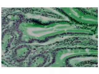
- Nucleic acid staining techniques are widely used in histology and microbiology to identify and visualize DNA and RNA within cells.
- These stains help differentiate cell types, study cell structures, and detect microorganisms.
- Stains that target DNA and RNA are critical for analyzing the cellular localization and distribution of genetic material.
- These special stains bind specifically to nucleic acids, producing distinct colors that can be visualized under a microscope.
Feulgen Reaction (Specific for DNA)
Principle
- The Feulgen reaction is one of the classic DNA-specific staining methods.
- The principle behind it is based on the fact that DNA contains aldehyde groups, which can be released by hydrolyzing the DNA with an acid (usually hydrochloric acid).
- This hydrolysis exposes aldehyde groups, allowing them to bind to Schiff reagent, forming a magenta or purple complex.
Materials and Reagents
- Tissue samples fixed in neutral-buffered formalin
- Hydrochloric acid (1N HCl) for hydrolysis
- Schiff reagent (freshly prepared or commercially available)
- Sodium metabisulfite or potassium metabisulfite solution to stabilize Schiff reaction (0.5-1%)
- Counterstain (optional): light green SF or nuclear fast red
- Deionized water
Procedure
- Fixation and Hydrolysis:
-
- Use 10% neutral-buffered formalin-fixed tissue sections.
- Place the slides in 1N HCl at 60°C for 8–12 minutes for controlled hydrolysis (this step liberates aldehyde groups).
- Application of Schiff Reagent:
-
- Rinse slides in distilled water to remove excess acid.
- Place the slides in Schiff reagent for 10-15 minutes until a pink-magenta color develops, indicating DNA presence.
- Stabilization with Metabisulfite Solution:
-
- Rinse in a metabisulfite solution to stop the reaction.
- Rinse under running tap water to stabilize the staining reaction fully.
- Counterstaining and Mounting:
-
- (Optional) Counterstain with light green or nuclear fast red for 1–2 minutes.
- Dehydrate in graded ethanol, clear in xylene, and mount with a coverslip.
Results and Interpretation
- Positive for DNA: Nuclei will appear bright magenta.
- Non-DNA elements Should remain unstained or lightly stained by the counterstain.
Applications
- Widely used in cytogenetics to measure DNA content in cells.
- Useful in cancer pathology for detecting abnormal DNA distributions and ploidy (DNA content) in malignant cells.
- Valuable in identifying cells undergoing mitosis or apoptosis based on DNA fragmentation patterns.
Methyl Green-Pyronin (MGP) Staining (Differentiates DNA and RNA)
Principle
- The MGP stain differentiates between DNA and RNA by using two dyes: methyl green (which binds to DNA, staining it green) and pyronin Y (which binds to RNA, staining it red).
- This differential staining is due to DNA and RNA’s structural and charge differences.
Materials and Reagents
- Tissue samples fixed in formalin or ethanol
- Methyl green solution (0.5% in acetate buffer, pH 4.2-4.5)
- Pyronin Y solution (0.5% in acetate buffer)
- Acetate buffer (pH 4.2-4.5)
- Distilled water
Procedure
- Preparation of Staining Solution:
-
- Mix methyl green and pyronin Y in acetate buffer to form the staining solution.
- Staining:
-
- Place slides in the methyl green-pyronin solution for 10–15 minutes.
- Rinsing and Mounting:
-
- Rinse slides with acetate buffer or distilled water to remove excess stain.
- Dehydrate, clear in xylene, and mount.
Results and Interpretation
- DNA-rich areas (nuclei): Stain green.
- RNA-rich areas (cytoplasm and nucleoli): Stain red or pink.
Applications
- Commonly used in immunology and hematology to differentiate between plasma cells (which contain high RNA content) and lymphocytes.
- Helps visualize DNA/RNA distribution in different cell types, which is useful for cancer diagnosis and cellular differentiation studies.
Acridine Orange (AO) Fluorescent Staining
Principle
- Acridine orange is a fluorescent stain that binds to nucleic acids.
- When excited under UV light, acridine orange emits green fluorescence when bound to DNA and red fluorescence when bound to RNA.
- This stain is especially useful for distinguishing live versus dead bacteria in microbial studies.
Materials and Reagents
- Acridine orange solution (0.01% in phosphate buffer, pH 6.5-7.0)
- Phosphate buffer (pH 6.5-7.0)
- Ethanol for fixation
Procedure
- Fixation:
-
- Fix cells or tissues in ethanol for optimal preservation of nucleic acids.
- Staining:
-
- Immerse slides in acridine orange solution for 5–10 minutes in a dark environment to prevent photobleaching.
- Rinsing:
-
- Rinse in phosphate buffer to remove excess stain.
- Observation:
-
- View immediately under a fluorescence microscope with UV excitation.
Results and Interpretation
- DNA: Emits green fluorescence.
- RNA: Emits orange to red fluorescence.
Applications
- Extensively used in microbiology for live-dead assays of bacterial samples.
- Applied in cytology to differentiate DNA from RNA in cells, useful in cell cycle studies and oncology.
Toluidine Blue Staining (Selective for RNA)
Principle
- Toluidine blue is a basic dye that binds to nucleic acids and produces a metachromatic effect, staining RNA-rich areas in a distinct color.
- This method is particularly useful for detecting ribosomes and RNA-heavy regions in the cytoplasm.
Materials and Reagents
- Toluidine blue solution (0.1% in sodium chloride or acetate buffer, pH 4.0-4.5)
- Distilled water for rinsing
- Sodium chloride or acetate buffer
Procedure
- Preparation of Solution:
-
- Dissolve toluidine blue in sodium chloride or acetate buffer to create the staining solution.
- Staining:
-
- Stain tissue sections with toluidine blue solution for 2–5 minutes.
- Rinsing and Mounting:
-
- Rinse in distilled water and cover slides with a coverslip.
Results and Interpretation
- RNA-rich areas (ribosomes, nucleoli): Appear deep blue to purple.
- Non-RNA areas: Show less intense staining or may remain clear.
Applications
- Used in histopathology to visualize mast cells, which stain intensely due to RNA content.
- Commonly applied for identifying bacteria or fungal elements in tissue samples based on their RNA-rich structures.
Applications Across Staining Techniques
Each nucleic acid stain offers unique visualization capabilities, enabling their applications in a wide range of fields:
- Feulgen Reaction:
-
- Essential for DNA quantification studies.
- Used in ploidy studies, cancer diagnosis, and cytogenetics.
- Methyl Green-Pyronin (MGP):
-
- Valuable in immunology and pathology for differentiating cell types based on nucleic acid content.
- Applied in lymphoma and leukemia diagnoses.
- Acridine Orange:
-
- Ideal for detecting live versus dead cells in microbial and environmental samples.
- Useful in cell cycle studies and apoptosis assays.
- Toluidine Blue:
-
- Applied in bacteriology and mycology for RNA-rich microbial identification.
- Useful for detecting mast cells and for general histology.