Introduction
-
Routine urine analysis is a commonly performed laboratory investigation used for screening and diagnosis of various diseases.
-
It is a simple, non-invasive, and cost-effective test.
-
Urine reflects the functional status of the kidneys, urinary tract, and metabolic processes of the body.
-
The test provides early information about renal, metabolic, hepatic, and systemic disorders.
-
Routine urine analysis consists of three main components:
-
Physical examination
-
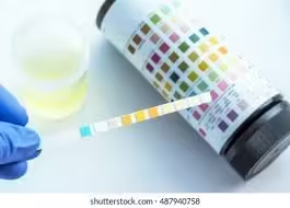
Chemical examination
-
Microscopic examination
-
-
Physical examination includes assessment of volume, color, appearance, and specific gravity.
-
Chemical examination detects substances such as protein, glucose, ketone bodies, bilirubin, and blood.
-
Microscopic examination helps identify cells, casts, crystals, and microorganisms.
-
It is widely used in general health check-ups and clinical evaluations.
-
Early detection of abnormalities through routine urine analysis assists in diagnosis, monitoring of disease, and assessment of treatment response.
Components of Urine
| Category | Component | Source / Formation | Normal Presence | Clinical Significance |
|---|---|---|---|---|
| Water | Water | Derived from plasma during glomerular filtration | 95–96% of urine | Reflects hydration status and renal concentrating ability |
| Organic components (Nitrogenous wastes) | Urea | Formed in liver from ammonia during protein metabolism | Major organic constituent | Increased in dehydration, renal failure; decreased in liver disease |
| Creatinine | Produced from muscle creatine phosphate breakdown | Constant daily excretion | Indicator of renal function (GFR) | |
| Uric acid | End product of purine metabolism | Present in moderate amount | Increased in gout, leukemia, high cell turnover | |
| Ammonia | Formed from glutamine in renal tubules | Present in small amount | Helps maintain acid–base balance | |
| Organic components (Non-nitrogenous) | Hippuric acid | Formed in liver from benzoic acid | Present | Increased after fruit intake or drug metabolism |
| Amino acids | Filtered and mostly reabsorbed | Traces only | Aminoaciduria in tubular disorders | |
| Glucose | Filtered and reabsorbed in PCT | Normally absent | Present in diabetes mellitus, renal glycosuria | |
| Ketone bodies | Formed during fat metabolism | Absent | Seen in diabetic ketoacidosis, starvation | |
| Bilirubin | Conjugated bilirubin from liver | Absent | Indicates obstructive or hepatic jaundice | |
| Urobilinogen | Formed from bilirubin in intestine | Trace amounts | Increased in hemolytic anemia; absent in obstructive jaundice | |
| Inorganic components (Electrolytes) | Sodium (Na⁺) | Dietary intake and renal regulation | Variable | Reflects fluid and electrolyte balance |
| Potassium (K⁺) | Regulated by kidneys and aldosterone | Variable | Altered in renal and adrenal disorders | |
| Chloride (Cl⁻) | Derived from dietary salt | Present | Altered in dehydration and acid–base disorders | |
| Phosphates | From protein and bone metabolism | Present | Buffering action; increased in renal disease | |
| Sulfates | From sulfur-containing amino acids | Present | Reflects protein catabolism | |
| Calcium | From bone and dietary sources | Small amount | Increased in hyperparathyroidism, renal stones | |
| Magnesium | From dietary sources | Small amount | Altered in renal disorders | |
| Pigments | Urochrome | Breakdown product of hemoglobin | Present | Responsible for normal yellow color |
| Urobilin | Oxidized form of urobilinogen | Present | Contributes to urine color | |
| Uroerythrin | Minor pigment | Trace | Gives reddish tint in concentrated urine | |
| Cellular elements (normally absent or minimal) | RBCs | Leakage from urinary tract | Absent | Hematuria in stones, tumors, glomerulonephritis |
| WBCs | From inflammation or infection | Absent | Indicates urinary tract infection | |
| Epithelial cells | Shed from urinary tract lining | Few | Increased in infections or tubular damage | |
| Casts | Formed in renal tubules | Absent | Specific types indicate renal pathology | |
| Crystals | Precipitated solutes | Occasional | Stone formation or metabolic disorders |
Specimen Requirements
| Parameter | Important Point |
|---|---|
| Type of specimen | Freshly voided urine |
| Preferred sample | Early morning urine |
| Collection method | Clean-catch midstream urine |
| Container | Clean, dry, wide-mouthed, leak-proof |
| Volume required | 10–20 mL |
| Time for examination | Within 1–2 hours of collection |
| Storage (if delayed) | Refrigeration recommended |
| Contamination | Avoid feces, menstrual blood, vaginal secretions |
| Labeling | Patient name, date, and time required |
| Unsuitable sample | Old or contaminated urine |
Physical Examination
Volume
Urine volume refers to the total amount of urine excreted in a given period, usually measured over 24 hours, and it reflects renal function and fluid balance.
Normal urine volume:
- The normal adult urine output is 1–2 liters per day (approximately 1500 mL) with normal fluid intake.
Abnormal urine volume:
-
Polyuria: Excessive urine output, usually more than 2.5–3 liters per day. It is commonly seen in diabetes mellitus, diabetes insipidus, excessive fluid intake, and use of diuretics.
-
Oliguria: Reduced urine output, less than 400 mL per day in adults. It may occur in dehydration, shock, acute renal failure, or severe diarrhea and vomiting.
-
Anuria: Complete or near-complete absence of urine, usually less than 100 mL per day, seen in severe renal failure or urinary tract obstruction.
-
Nocturia: Increased frequency of urination at night, commonly associated with diabetes mellitus, urinary tract infections, cardiac failure, and prostatic enlargement.
| Parameter | Description / Definition | Clinical Significance |
|---|---|---|
| Normal urine volume | 1–2 liters/day (≈1500 mL) | Indicates normal renal function and hydration |
| Polyuria | >2.5–3 liters/day | Diabetes mellitus, diabetes insipidus, diuretics, excess fluid intake |
| Oliguria | <400 mL/day | Dehydration, shock, acute renal failure |
| Anuria | <100 mL/day | Severe renal failure, urinary tract obstruction |
| Nocturia | Increased urine output at night | Diabetes mellitus, cardiac failure, UTI, prostatic enlargement |
Colour
Urine colour is a key indicator of hydration and can also reflect the presence of certain substances or diseases.
Normal colour:
- The normal colour of urine ranges from pale yellow to amber due to the presence of the pigment urochrome.
- The intensity of colour depends on the concentration of urine.
- Light-coloured urine indicates good hydration, whereas dark yellow urine suggests dehydration.
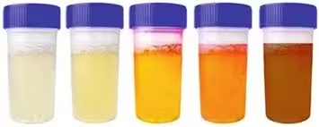
Abnormal colours:
-
Dark yellow or amber: Indicates concentrated urine, commonly seen in dehydration or excessive sweating.
-
Red or pink: Usually due to the presence of blood (hematuria); it may also occur after intake of certain foods such as beetroot or drugs like rifampin.
-
Brown: Suggests liver disease or the presence of bile pigments, as seen in jaundice; some medications may also cause brown urine.
-
Orange: May be due to dehydration, intake of carotene-rich foods, or drugs such as phenazopyridine.
-
Blue or green: Rare; can occur in bacterial infections such as Pseudomonas or due to medications like methylene blue.
-
Milky or cloudy: Indicates the presence of pus cells, bacteria, or crystals and is suggestive of urinary tract infection or renal stone disease.
| Urine color | Causing substance | Occurrence / Clinical conditions |
|---|---|---|
| Yellow to colorless | Dilute urine | Increased diuresis due to excessive fluid intake, diuretic drugs, diabetes mellitus, diabetes insipidus, polyuric phase of renal failure |
| Brown | Bilirubin | Diseases of liver and biliary tract |
| Green-brown | Biliverdin (formed from oxidation of bilirubin on exposure to air; old urine) | Diseases of liver and biliary tract |
| Yellow-orange | Riboflavin, carotenes | Exogenous intake (vitamins, carotene-rich foods) |
| Meat red (without turbidity) | Hemoglobin, myoglobin, porphyrins, beetroot pigments | Intravascular hemolysis, burns, muscle necrosis, muscle inflammation, porphyrias, exogenous intake |
| Meat red (with turbidity) | Blood (RBCs) | Macroscopic hematuria due to diseases of kidney and urinary tract, disorders of hemostasis, bleeding into urinary tract |
| Dark brown (turns black on standing) | Melanin, homogentisic acid | Melanoma, alkaptonuria |
| Light red | Urates | Hyperuricosuria |
Turbidity
Turbidity refers to the clarity or cloudiness of urine and is assessed by visual inspection.
Normal urine:
- Freshly passed urine is clear and transparent. On standing, slight cloudiness may develop due to precipitation of salts.
Abnormal turbidity:
-
Cloudy urine: May be due to the presence of pus cells (leukocytes), red blood cells, epithelial cells, bacteria, or mucus, commonly seen in urinary tract infections.
-
Milky urine: Suggests the presence of pus (pyuria), chyle (chyluria), or excess phosphates.
-
Smoky appearance: Often caused by red blood cells and is characteristic of glomerular disorders.
-
Turbidity on standing: Due to precipitation of urates in acidic urine or phosphates in alkaline urine.
| Appearance | Cause | Clinical Significance |
|---|---|---|
| Clear | Normal urine | Indicates absence of suspended particles |
| Slightly cloudy on standing | Precipitation of salts | Normal finding |
| Cloudy | Pus cells, RBCs, epithelial cells, bacteria, mucus | Urinary tract infection |
| Milky | Pus, chyle, phosphates | Pyuria, chyluria |
| Smoky | Red blood cells | Glomerular disorders |
| Turbidity on standing | Urates (acidic urine) or phosphates (alkaline urine) | Crystalluria |
Odour
Normal odour:
- Freshly voided urine has a mild, characteristic odour due to the presence of metabolic waste products such as urea.
Foul or strong odour:
-
Urinary tract infection (UTI): A strong, foul-smelling odour is commonly associated with bacterial infection due to decomposition of urea by bacteria.
-
Dehydration: Highly concentrated urine produces a stronger odour because of increased concentration of waste products.
-
Ketosis (fruity odour): In conditions such as diabetic ketoacidosis or prolonged fasting, increased fat metabolism leads to ketone body formation, giving urine a fruity or sweet smell.
-
Food and medications: Certain foods like asparagus and garlic, and drugs such as antibiotics, can cause a strong or unusual urine odour.
| Odour | Cause | Clinical Significance |
|---|---|---|
| Aromatic | Normal urine | Freshly voided normal urine |
| Ammoniacal | Bacterial decomposition of urea | Old urine, urinary tract infection |
| Fruity / sweet | Presence of ketone bodies | Diabetes mellitus, diabetic ketoacidosis |
| Foul-smelling | Bacterial infection | Urinary tract infection |
| Fishy | Trimethylamine | Metabolic disorders |
| Mousy | Phenylalanine metabolites | Phenylketonuria |
Specific Gravity
Specific gravity is a measure of the concentration of urine and reflects the kidney’s ability to concentrate or dilute urine.
Normal specific gravity:
- The normal range of urine specific gravity is 1.015–1.025 (may vary between 1.005–1.030 depending on fluid intake).
Increased specific gravity:
- An increased specific gravity indicates concentrated urine and is commonly seen in dehydration, excessive sweating, diarrhea, vomiting, and conditions such as diabetes mellitus (due to glucose in urine).
Decreased specific gravity:
- A decreased specific gravity indicates dilute urine and occurs in excessive fluid intake, diabetes insipidus, chronic renal failure, and conditions where the kidneys lose their concentrating ability.
Fixed specific gravity (Isosthenuria):
- A constant specific gravity around 1.010 suggests loss of renal concentrating and diluting power, commonly seen in chronic renal disease.
| Term | Value of relative specific gravity | Causes / Clinical conditions |
|---|---|---|
| Eusthenuria | 1.020 – 1.040 | Normal concentrating ability of kidneys |
| Hypersthenuria | ↑ > 1.040 | Dehydration, glucosuria, proteinuria |
| Hyposthenuria | ↓ < 1.020 | Diabetes insipidus, hyperhydration, renal failure, use of diuretic drugs |
| Isosthenuria | = 1.010 | Severe kidney damage with loss of concentrating and diluting ability |
pH
The normal range of urine pH is 4.5 to 8.0, with an average value of about 6.0, indicating that urine is usually slightly acidic.
Factors Affecting Urine pH
Diet:
-
A high-protein diet (meat, eggs) produces acidic urine due to increased acid load, resulting in a lower pH.
-
A vegetarian diet and high intake of fruits and vegetables produce alkaline urine, resulting in a higher pH.
Medications:
-
Drugs such as sodium bicarbonate make urine more alkaline.
-
Drugs like ammonium chloride and some acidifying agents lower urine pH.
Metabolic and respiratory conditions:
-
Metabolic or respiratory acidosis leads to acidic urine as the kidneys excrete excess hydrogen ions.
-
Metabolic or respiratory alkalosis results in alkaline urine due to reduced hydrogen ion excretion.
Clinical Significance of Urine pH
Acidic urine (pH < 5.5):
-
Seen in metabolic acidosis, diabetic ketoacidosis, starvation, and high animal-protein intake.
-
Associated with formation of uric acid and cystine renal stones.
Alkaline urine (pH > 7.5):
-
Suggests urinary tract infection caused by urease-producing bacteria, which convert urea into ammonia.
-
Seen in metabolic alkalosis, vegetarian diet, and after intake of alkaline drugs.
-
Favors formation of struvite (magnesium ammonium phosphate) stones.
| Urine pH | Range / Value | Causes / Conditions |
|---|---|---|
| Normal | 4.5 – 8.0 (average ≈ 6.0) | Mixed diet; normal renal acid–base regulation |
| Acidic urine | < 5.5 | High-protein diet, metabolic acidosis, diabetic ketoacidosis, starvation, fever |
| Alkaline urine | > 7.5 | Vegetarian diet, metabolic alkalosis, vomiting, post-prandial alkaline tide |
| Alkaline urine (pathological) | > 7.5 | Urinary tract infection with urease-producing bacteria |
| Drug-induced acidic urine | ↓ pH | Ammonium chloride, ascorbic acid |
| Drug-induced alkaline urine | ↑ pH | Sodium bicarbonate, acetazolamide |
| Stone association (acidic) | — | Uric acid and cystine stones |
| Stone association (alkaline) | — | Struvite (magnesium ammonium phosphate) stones |
MCQs
1. Routine urine analysis is primarily used for:
A. Treatment of diseases
B. Screening and diagnosis of diseases
C. Surgical evaluation
D. Genetic counseling
Answer: B
2. Routine urine examination is considered:
A. Invasive and costly
B. Invasive and cheap
C. Non-invasive and cost-effective
D. Non-invasive but expensive
Answer: C
3. Urine reflects the functional status of:
A. Liver only
B. Heart only
C. Kidneys, urinary tract, and metabolism
D. Lungs
Answer: C
4. Routine urine analysis provides early information about:
A. Only renal disorders
B. Only metabolic disorders
C. Renal, metabolic, hepatic, and systemic disorders
D. Neurological disorders
Answer: C
5. Routine urine analysis consists of how many main components?
A. Two
B. Three
C. Four
D. Five
Answer: B
6. Which of the following is NOT a component of routine urine analysis?
A. Physical examination
B. Chemical examination
C. Microscopic examination
D. Radiological examination
Answer: D
7. Physical examination of urine includes assessment of:
A. Protein and glucose
B. Volume, color, appearance, specific gravity
C. Cells and casts
D. Electrolytes
Answer: B
8. Chemical examination of urine detects:
A. Cells and crystals
B. Bacteria only
C. Protein, glucose, ketones, bilirubin, blood
D. Casts only
Answer: C
9. Microscopic examination of urine helps identify:
A. Electrolytes
B. Pigments
C. Cells, casts, crystals, microorganisms
D. Hormones
Answer: C
10. Routine urine analysis is commonly used in:
A. Emergency surgery only
B. Research laboratories only
C. General health check-ups
D. Veterinary practice only
Answer: C
MCQs on Components of Urine
11. The major component of urine is:
A. Urea
B. Water
C. Creatinine
D. Electrolytes
Answer: B
12. Water constitutes approximately what percentage of urine?
A. 50–60%
B. 70–80%
C. 85–90%
D. 95–96%
Answer: D
13. The major nitrogenous waste product in urine is:
A. Creatinine
B. Uric acid
C. Urea
D. Ammonia
Answer: C
14. Urea is formed in the:
A. Kidney
B. Muscle
C. Liver
D. Intestine
Answer: C
15. Creatinine excretion is important because it reflects:
A. Liver function
B. Muscle mass
C. Renal function (GFR)
D. Hydration status
Answer: C
16. Increased uric acid in urine is seen in:
A. Diabetes insipidus
B. Gout and leukemia
C. Liver failure
D. Renal tuberculosis
Answer: B
17. Ammonia in urine helps maintain:
A. Osmotic balance
B. Electrolyte balance
C. Acid–base balance
D. Blood pressure
Answer: C
18. Glucose is normally:
A. Present in large amounts
B. Present in moderate amounts
C. Absent in urine
D. Always present
Answer: C
19. Presence of glucose in urine indicates:
A. Liver disease
B. Diabetes mellitus
C. Gout
D. UTI
Answer: B
20. Ketone bodies in urine are seen in:
A. Liver cirrhosis
B. Nephrotic syndrome
C. Diabetic ketoacidosis
D. UTI
Answer: C
MCQs on Specimen Requirements
21. The preferred urine sample for routine examination is:
A. Random urine
B. Post-meal urine
C. Early morning urine
D. Evening urine
Answer: C
22. The recommended urine collection method is:
A. First stream urine
B. Clean-catch midstream urine
C. Catheterized urine
D. Suprapubic aspirate
Answer: B
23. Minimum volume of urine required is:
A. 2–5 mL
B. 5–10 mL
C. 10–20 mL
D. 50 mL
Answer: C
24. Urine should ideally be examined within:
A. 10 minutes
B. 30 minutes
C. 1–2 hours
D. 24 hours
Answer: C
25. Delay in urine examination can be minimized by:
A. Heating the sample
B. Adding glucose
C. Refrigeration
D. Shaking the sample
Answer: C
MCQs on Urine Volume
26. Normal adult urine output per day is:
A. 200–400 mL
B. 500–800 mL
C. 1–2 liters
D. 3–4 liters
Answer: C
27. Polyuria is defined as urine output:
A. <400 mL/day
B. <100 mL/day
C. >2.5–3 liters/day
D. >1 liter/day
Answer: C
28. Oliguria refers to urine output:
A. >3 liters/day
B. <400 mL/day
C. <100 mL/day
D. Normal output
Answer: B
29. Anuria is urine output:
A. <500 mL/day
B. <200 mL/day
C. <100 mL/day
D. <50 mL/day
Answer: C
30. Nocturia is commonly seen in:
A. Diabetes mellitus
B. Cardiac failure
C. Prostatic enlargement
D. All of the above
Answer: D
MCQs on Colour, Turbidity, Odour, SG, pH
31. Normal urine colour is due to:
A. Bilirubin
B. Hemoglobin
C. Urochrome
D. Urobilinogen
Answer: C
32. Red urine without turbidity suggests:
A. Hematuria
B. Hemoglobinuria
C. Pyuria
D. Phosphaturia
Answer: B
33. Milky urine is commonly due to:
A. Glucose
B. Protein
C. Pus or chyle
D. Ketones
Answer: C
34. Smoky urine appearance is typical of:
A. UTI
B. Glomerular disease
C. Diabetes mellitus
D. Liver disease
Answer: B
35. Ammoniacal odour of urine suggests:
A. Fresh urine
B. Ketosis
C. Old urine or UTI
D. Liver disease
Answer: C
36. Fruity odour of urine is due to:
A. Glucose
B. Ketone bodies
C. Ammonia
D. Urea
Answer: B
37. Normal urine specific gravity range is:
A. 1.000–1.005
B. 1.005–1.030
C. 1.040–1.060
D. 1.060–1.080
Answer: B
38. Isosthenuria indicates:
A. Dehydration
B. Diabetes mellitus
C. Severe kidney damage
D. Liver disease
Answer: C
39. Hyposthenuria is seen in:
A. Dehydration
B. Diabetes insipidus
C. Proteinuria
D. Glucosuria
Answer: B
40. Normal urine pH is:
A. Always alkaline
B. Always acidic
C. 4.5–8.0
D. 7.5–9.0
Answer: C
41. Acidic urine favors formation of:
A. Struvite stones
B. Calcium stones
C. Uric acid stones
D. Phosphate stones
Answer: C
42. Alkaline urine is commonly seen in:
A. Starvation
B. High-protein diet
C. UTI with urease-producing bacteria
D. Diabetic ketoacidosis
Answer: C
43. Vegetarian diet usually produces:
A. Acidic urine
B. Neutral urine
C. Alkaline urine
D. No change in urine pH
Answer: C
44. Sodium bicarbonate intake causes urine to become:
A. Acidic
B. Neutral
C. Alkaline
D. Colorless
Answer: C
45. Urobilinogen is increased in:
A. Obstructive jaundice
B. Hemolytic anemia
C. Renal failure
D. UTI
Answer: B
46. Presence of RBCs in urine is termed:
A. Pyuria
B. Hematuria
C. Proteinuria
D. Glycosuria
Answer: B
47. Presence of WBCs in urine indicates:
A. Diabetes mellitus
B. Renal stones
C. Urinary tract infection
D. Liver disease
Answer: C
48. Casts are formed in:
A. Bladder
B. Ureter
C. Renal tubules
D. Urethra
Answer: C
49. Uroerythrin gives urine a:
A. Yellow color
B. Brown color
C. Reddish tint
D. Green color
Answer: C
50. Early detection through routine urine analysis helps in:
A. Only diagnosis
B. Only treatment
C. Diagnosis, monitoring, and treatment response
D. Surgery planning
Answer: C