Introduction
- The preparation of tissue sections is an essential step in histopathology.
- Different techniques are used depending on whether we want to study gross tissue architecture, cellular details, cytology, enzyme activity, or ultrastructure.
Paraffin Wax Embedding Method
-
Most widely used method in diagnostic histopathology.
-
Produces thin, high-quality sections suitable for staining and long-term storage.
Steps:
-
Fixation
-
Prevents autolysis and putrefaction.
-
Common fixative: 10% Neutral Buffered Formalin (NBF).
-
Fixation time: 6–48 hours (depending on tissue size).
-
-
Dehydration
-
Tissue water is removed using increasing grades of alcohol (70%, 80%, 90%, 100%).
-
-
Clearing
-
Replaces alcohol with a clearing agent (xylene, chloroform, or toluene).
-
Makes tissue transparent and miscible with paraffin.
-
-
Infiltration
-
Tissue is impregnated with molten paraffin wax (56–60°C).
-
-
Embedding
-
Tissue is oriented properly in a wax block.
-
Block is cooled and solidified.
-
-
Section Cutting
-
Done with a rotary microtome.
-
Typical thickness: 3–5 µm for light microscopy.
-
-
Staining
-
Routine: Hematoxylin & Eosin (H&E).
-
Special stains: PAS, Masson’s Trichrome, Reticulin, etc.
-
-
Mounting
-
Sections are mounted with DPX or Canada balsam and covered with a coverslip.
-
✔ Advantages: Excellent preservation of morphology, permanent slides.
✘ Disadvantages: Slow process (12–24 hrs), lipids and some antigens may be lost.
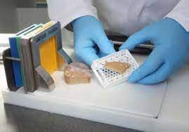
Frozen Section Method
-
Used when rapid intraoperative diagnosis is required (oncology, margins of excision).
-
Preserves lipids, enzymes, and antigens better than paraffin.
Steps:
-
Fresh tissue rapidly frozen using liquid nitrogen, isopentane, or cryostat refrigerant.
-
Sectioned using a cryostat microtome at −20°C.
-
Sections mounted on glass slides.
-
Stained quickly with rapid H&E, Oil Red O (for fat), or enzyme histochemistry.
✔ Advantages: Very fast (results in 10–15 min), useful for enzyme/lipid studies.
✘ Disadvantages: Poorer section quality, cannot be preserved for long periods.
Cell Block Technique
-
Special method for cytology specimens (body fluids, FNAC aspirates).
-
Converts dispersed cells into a tissue-like block.
Steps:
-
Centrifugation of sample.
-
Formation of a cell clot (by adding plasma + thrombin, or by natural sedimentation).
-
Fixed in formalin and processed like paraffin blocks.
-
Sections cut and stained.
✔ Advantages: Preserves cell architecture, multiple stains possible.
✘ Disadvantages: Requires good cellularity.
Resin Embedding Method
-
Used in electron microscopy (EM) and sometimes for very hard tissues (e.g., bone, teeth).
Steps:
-
Fixation: Glutaraldehyde (primary) + Osmium tetroxide (secondary).
-
Dehydration: Acetone or alcohol.
-
Infiltration: With epoxy resins (Epon, Araldite) or acrylic resins.
-
Polymerization: Resin block hardened by heat or UV light.
-
Ultramicrotome used for sectioning (50–100 nm thickness).
-
Staining with heavy metals (uranyl acetate, lead citrate) for EM contrast.
✔ Advantages: Preserves ultrastructure of organelles.
✘ Disadvantages: Expensive, requires specialized setup.
Cell Smear / Impression Cytology
-
Used for cytology and quick diagnosis.
-
Types:
-
Touch Preparation (Imprint Smear): Tissue surface pressed onto a slide.
-
Crush Smear: Tissue gently crushed between two slides.
-
Scrape Smear: Tissue scraped and spread on slide.
-
✔ Advantages: Simple, quick, inexpensive.
✘ Disadvantages: No tissue architecture preserved, only cytological details.
Whole Mount Sections
-
Small specimens (embryos, insects, worms) are embedded and sectioned in entirety.
-
Useful in developmental biology, parasitology, embryology.
✔ Advantages: Study of the entire structure.
✘ Disadvantages: Only small specimens can be used.
Free-Hand Section
-
Oldest method, still used in botany labs and basic teaching.
-
Fresh or fixed tissue cut manually with a razor blade.
✔ Advantages: No equipment required, very quick.
✘ Disadvantages: Uneven sections, poor detail.
Special Embedding Techniques
-
Celloidin Embedding
-
Uses celloidin (a form of nitrocellulose).
-
Good for brain, eye, and delicate tissues.
-
Provides support, avoids shrinkage.
-
-
Double Embedding (Paraffin + Celloidin)
-
Used for hard tissues like bone.
-
-
Gelatin Embedding
-
For frozen sections, particularly muscle and nerve tissues.
-
Comparison Table
| Method | Section Thickness | Time Required | Preservation | Best For |
|---|---|---|---|---|
| Paraffin Wax | 3–5 µm | 12–24 hrs | Good | Routine histology |
| Frozen Section | 8–12 µm | 10–20 min | Moderate | Rapid diagnosis, lipids |
| Resin Embedding | 50–100 nm | 2–7 days | Excellent | Electron microscopy |
| Cell Block | 3–5 µm | 1 day | Good | Cytology samples |
| Smear/Imprint | — | Immediate | Fair | Cytology |
| Free-Hand Section | 20–100 µm | Immediate | Poor | Teaching, botany |