Introduction
Romanowsky dyes are a group of polychromatic staining techniques that are fundamental to diagnostic haematology. These stains are primarily used for:
-
Peripheral blood smear examination
-
Bone marrow smear evaluation
-
Detection of haemoparasites
-
Cytogenetic studies
The major strength of Romanowsky stains lies in their ability to produce a wide range of colours using a combination of acidic and basic dyes. This unique staining reaction, known as the Romanowsky effect, allows clear differentiation between nucleus, cytoplasm, and granules of various blood cells.
In routine laboratory practice, Romanowsky stains help in:
-
Performing Differential Leukocyte Count (DLC)
-
Identifying abnormal WBCs (blasts, atypical lymphocytes)
-
Evaluating RBC morphology
-
Assessing platelet disorders
-
Detecting infectious agents
Without Romanowsky staining, microscopic evaluation of blood cells would lack clarity and diagnostic precision.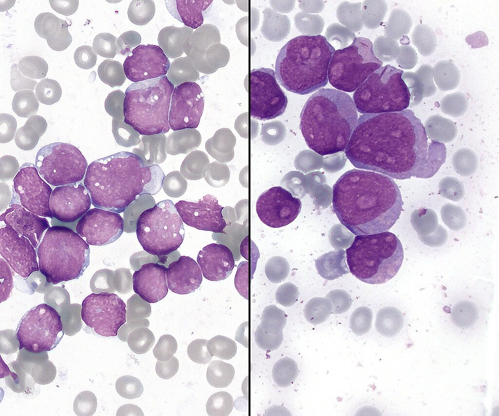
Composition of Romanowsky Dyes
Romanowsky stains are composed of a mixture of:
A. Basic Dye (Cationic Dye)
1. Methylene Blue
-
A thiazine dye.
-
Positively charged.
-
Binds to negatively charged (acidic) cellular components.
2. Azure B
-
Oxidation product of methylene blue.
-
Responsible for the characteristic purple nuclear staining.
-
Plays a key role in the Romanowsky effect.
Structures Stained by Basic Dyes:
-
DNA (nucleus)
-
RNA (ribosomes)
-
Rough endoplasmic reticulum
-
Basophilic granules
-
Platelet granules
These structures appear:
-
Blue
-
Dark blue
-
Purple
B. Acidic Dye (Anionic Dye)
Eosin Y
-
Negatively charged dye.
-
Binds to positively charged (basic) proteins.
Structures Stained by Eosin:
-
Haemoglobin
-
Cytoplasmic proteins
-
Eosinophilic granules
These structures appear:
-
Pink
-
Orange
-
Red
Romanowsky Effect
The Romanowsky effect occurs due to interaction between:
-
Azure B (basic dye)
-
Eosin (acidic dye)
This interaction forms a complex that stains:
-
Chromatin → Purple
-
Neutrophil granules → Lilac
-
Cytoplasm → Variable shades
This polychromasia makes Romanowsky stains superior to simple single-dye stains.
Types of Romanowsky Stains
Leishman Stain:
Composition
-
Methylene blue
-
Eosin
-
Methanol (acts as fixative)
Special Feature
-
Does not require separate fixation step.
-
Methanol fixes and stains simultaneously.
Detailed Microscopic Appearance
-
RBCs → Pink to salmon colour
-
Neutrophils → Multi-lobed nucleus (purple), lilac granules
-
Eosinophils → Bilobed nucleus, bright red-orange granules
-
Basophils → Dark blue-black granules
-
Lymphocytes → Dark purple nucleus, sky-blue cytoplasm
-
Monocytes → Kidney-shaped nucleus, grey-blue cytoplasm
Advantages
-
Rapid staining
-
Good nuclear detail
-
Suitable for routine DLC

Wright’s Stain:
Composition
-
Methylene blue
-
Eosin
-
Methanol
Characteristics
-
Commonly used in automated laboratories
-
Slightly faster than Giemsa
Detailed Staining Features
-
RBCs → Uniform pink
-
Neutrophils → Pink-lilac granules
-
Eosinophils → Intense orange granules
-
Basophils → Deep violet granules
-
Platelets → Purple granules
Clinical Importance
-
Routine blood smear examination
-
Identification of toxic granulation
-
Detection of abnormal leukocytes
Significance: Primarily used in haematology for routine blood smear examination.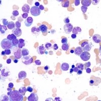
Giemsa Stain:
Composition
-
Azure B
-
Methylene blue
-
Eosin
-
Glycerol
Special Features
-
Provides superior chromatin detail
-
Best stain for parasite detection
Appearance
-
RBCs → Pale pink
-
Chromatin → Dark purple
-
Neutrophils → Fine lilac granules
-
Parasites → Distinct blue cytoplasm with red chromatin dots
Role in Malaria
-
Ring form → Blue ring with red chromatin
-
Schizont → Multiple merozoites
-
Gametocyte → Crescent shape (P. falciparum)
Role in Cytogenetics
-
Used in G-banding
-
Differentiates light and dark chromosomal bands
-
Detects chromosomal abnormalities
May-Grünwald Stain:
Composition: Like Wright’s stain, eosin and methylene blue are the main components.
Use: Commonly combined with Giemsa for a combined stain (May-Grünwald-Giemsa) for better resolution in haematological studies.
Appearance:
- Eosinophilic components appear orange-red.
- Basophilic components appear blue to purple.
Significance: Frequently used for blood smears, providing high-quality cellular detail.
Principle of Romanowsky Staining
The principle of Romanowsky staining is based on the differential affinity of the dyes for various cellular components, resulting in contrasting colours that allow easy identification and differentiation of blood cells:
Acidic (basophilic) structures, like DNA and RNA, are stained by the basic dye (methylene blue or azure B) and appear blue to purple.
Examples: Nucleus, ribosomes, granules in basophils.
Basic (eosinophilic or acidophilic) structures, like haemoglobin and cytoplasmic proteins, are stained by the acidic dye (eosin) and appear pink to red.
Examples: Cytoplasm, granules in eosinophils, and haemoglobin in red blood cells.
Mechanism of Staining
Fixation:
The blood smear is air-dried and fixed using Methanol (part of the stain in many Romanowsky variants). This preserves the cell morphology.
Staining:
The smear is flooded with the stain, allowing the acidic and basic dyes to bind to the cellular components.
Differentiation:
The slide is rinsed with buffer solution, allowing the excess dye to be removed while leaving the bound dye, resulting in differential staining of various cells.
Microscopic Examination:
Once dried, the slide is observed under a microscope, where different cells and their components show contrasting colours based on their affinity for the dyes.
Applications of Romanowsky Stains
Peripheral Blood Smear Examination:
Romanowsky stains are primarily used for examining peripheral blood smears to:
- Identify different types of white blood cells (WBCs), such as neutrophils, lymphocytes, monocytes, eosinophils, and basophils.
- Evaluate the morphology of red blood cells (RBCs) to detect abnormalities such as anisocytosis, poikilocytosis, and changes in haemoglobin concentration.
- Examine platelet morphology and number.
Bone Marrow Examination:
Bone marrow smears stained with Romanowsky dyes are crucial for:
- Diagnosing haematological disorders such as leukaemia, lymphoma, aplastic anaemia, and myeloproliferative disorders.
- Assessing the cellularity and development of various blood cell lineages.
Detection of Parasitic Infections:
Romanowsky stains, particularly Giemsa stains, are widely used to detect blood-borne parasites:
- Malaria parasites (Plasmodium species) show characteristic appearances, with the ring forms, schizonts, and gametocytes easily identified.
- Other parasites like Trypanosoma (causing sleeping sickness) and Leishmania (causing leishmaniasis) can also be detected.
Cytogenetics (G-Banding):
Giemsa stain is used in G-banding for karyotyping:
- This technique highlights chromosomal regions, allowing the detection of chromosomal abnormalities like translocations, deletions, duplications, and inversions.
Platelet Morphology:
Romanowsky stains are also used to evaluate platelets, helping diagnose thrombocytopenia or platelet function disorders.
Advantages of Romanowsky Stains
Differentiation of Cell Types:
It provides excellent contrast between different types of cells, allowing clear differentiation of WBCs, RBCs, and platelets.
Detection of Cell Abnormalities:
Allows visualization of abnormal cell morphology, essential in diagnosing conditions like leukaemia, anaemia, and infections.
Parasite Detection:
Especially useful for detecting blood parasites, such as malaria, with Giemsa stain being the gold standard.
Limitations
Artifact formation: Poor staining technique can result in artifacts that may be confused with cellular components.
Time-consuming: Staining can be time-intensive, especially when multiple smears are stained simultaneously.
Expertise required: Accurate interpretation of stained slides requires experience, particularly distinguishing between normal and abnormal cells.
MCQs
-
Romanowsky stains are primarily used for staining:
A. Tissue sections
B. Blood smears
C. Urine samples
D. CSF samples
Answer: B -
The Romanowsky effect is produced by interaction between:
A. Methanol and buffer
B. Eosin and azure B
C. Water and alcohol
D. Haemoglobin and DNA
Answer: B -
The basic dye in Romanowsky stain is:
A. Eosin
B. Haematoxylin
C. Methylene blue
D. Safranin
Answer: C -
Eosin is a:
A. Basic dye
B. Neutral dye
C. Acidic dye
D. Fluorescent dye
Answer: C -
DNA is stained by:
A. Eosin
B. Azure B
C. Methanol
D. Alcohol
Answer: B -
The optimal pH for Romanowsky staining is:
A. 4.5
B. 5.2
C. 6.8
D. 8.5
Answer: C -
In Romanowsky staining, RBCs appear:
A. Blue
B. Green
C. Pink
D. Black
Answer: C -
Neutrophil granules typically appear:
A. Bright red
B. Pink to lilac
C. Deep blue
D. Brown
Answer: B -
Eosinophil granules stain:
A. Blue
B. Orange-red
C. Black
D. Green
Answer: B -
Basophil granules appear:
A. Pale pink
B. Dark purple
C. Yellow
D. Grey
Answer: B -
Methanol in Leishman stain acts as:
A. Buffer
B. Fixative
C. Mordant
D. Decolorizer
Answer: B -
Which stain is considered gold standard for malaria detection?
A. Wright’s stain
B. Leishman stain
C. Giemsa stain
D. Gram stain
Answer: C -
Giemsa stain is also used in:
A. Gram staining
B. Acid-fast staining
C. G-banding of chromosomes
D. Endospore staining
Answer: C -
The purple color of nucleus is due to binding of:
A. Eosin
B. Methanol
C. Azure B
D. Glycerol
Answer: C -
Lymphocyte cytoplasm appears:
A. Light blue
B. Dark green
C. Yellow
D. Red
Answer: A -
Which stain combines May-Grünwald and Giemsa?
A. Wright-Giemsa
B. MGG stain
C. PAS
D. H&E
Answer: B -
Excessively blue staining indicates:
A. Acidic pH
B. Alkaline pH
C. Proper staining
D. Over-fixation
Answer: B -
Excessively red staining indicates:
A. Acidic pH
B. Alkaline pH
C. Thick smear
D. Proper washing
Answer: A -
Platelets appear:
A. Red discs
B. Purple granules
C. Green bodies
D. Transparent
Answer: B -
Which parasite is best visualized by Giemsa stain?
A. Taenia
B. Plasmodium
C. Ascaris
D. Hookworm
Answer: B -
The fixation step prevents:
A. Cell multiplication
B. Cell distortion
C. Dye binding
D. RBC formation
Answer: B -
Romanowsky stains are examples of:
A. Simple stains
B. Differential stains
C. Negative stains
D. Fluorescent stains
Answer: B -
The cytoplasm of monocytes appears:
A. Grey-blue
B. Orange
C. Bright red
D. Black
Answer: A -
Toxic granulation is seen in:
A. RBCs
B. Neutrophils
C. Platelets
D. Lymphocytes
Answer: B -
The major acidic dye in Romanowsky stains is:
A. Safranin
B. Eosin
C. Crystal violet
D. Malachite green
Answer: B -
Bone marrow smears are commonly stained using:
A. Gram stain
B. Ziehl-Neelsen stain
C. Romanowsky stain
D. India ink
Answer: C -
The Romanowsky effect produces:
A. Monochromatic staining
B. Polychromatic staining
C. No staining
D. Fluorescent staining
Answer: B -
Schizonts of malaria are characterized by:
A. Single nucleus
B. Multiple merozoites
C. Flagella
D. Spores
Answer: B -
Crescent-shaped gametocytes are seen in:
A. P. vivax
B. P. malariae
C. P. falciparum
D. P. ovale
Answer: C -
A thick smear may result in:
A. Clear morphology
B. Artifact formation
C. No staining
D. Perfect differentiation
Answer: B -
The main structure responsible for purple chromatin staining is:
A. Lipids
B. Carbohydrates
C. DNA
D. Water
Answer: C -
Which cell shows bilobed nucleus and orange granules?
A. Neutrophil
B. Basophil
C. Eosinophil
D. Monocyte
Answer: C -
Basophils are identified by:
A. Pink cytoplasm
B. Dark blue granules
C. Red nucleus
D. Yellow granules
Answer: B -
The staining quality depends on:
A. Temperature only
B. pH only
C. pH and timing
D. Slide color
Answer: C -
RBC anisocytosis refers to variation in:
A. Shape
B. Color
C. Size
D. Number
Answer: C -
Poikilocytosis refers to variation in:
A. Size
B. Shape
C. Color
D. Count
Answer: B -
Hypochromia indicates:
A. Increased hemoglobin
B. Decreased hemoglobin
C. Increased platelets
D. Increased WBCs
Answer: B -
Azure B is derived from:
A. Eosin
B. Methanol
C. Methylene blue
D. Glycerol
Answer: C -
Methanol fixation occurs for approximately:
A. 5 seconds
B. 1 minute
C. 10 minutes
D. 1 hour
Answer: B -
The oil immersion objective used is:
A. 10×
B. 40×
C. 60×
D. 100×
Answer: D -
Romanowsky stains are widely used in:
A. Dermatology only
B. Haematology
C. Dentistry
D. Orthopedics
Answer: B -
Myeloblasts show:
A. No nucleus
B. Prominent nucleoli
C. Orange cytoplasm
D. No cytoplasm
Answer: B -
Platelet clumping may cause:
A. False high platelet count
B. False low platelet count
C. Leukocytosis
D. Anemia
Answer: B -
Which component stains haemoglobin?
A. Azure B
B. Eosin
C. Methanol
D. Buffer
Answer: B -
The kidney-shaped nucleus is typical of:
A. Monocyte
B. Neutrophil
C. Eosinophil
D. Basophil
Answer: A -
Romanowsky stains are not suitable for:
A. DLC
B. Parasite detection
C. Blood grouping
D. Bone marrow study
Answer: C -
Improper washing leads to:
A. Clear slide
B. Artifact formation
C. Better differentiation
D. No effect
Answer: B -
The cytoplasm of RBC lacks:
A. Haemoglobin
B. DNA
C. Protein
D. Iron
Answer: B -
The major diagnostic advantage of Romanowsky stains is:
A. Fluorescence
B. Color differentiation
C. Smell
D. Thickness
Answer: B -
Romanowsky stains are indispensable in diagnosing:
A. Fractures
B. Diabetes
C. Leukaemia
D. Hypertension
Answer: C